Categories
Blog

Exploring the Potential of Cornea Transplants: A Glimpse into a Promising Future

Exploring the Potential of Cornea Transplants: A Glimpse into a Promising Future

Cornea-Transplants

The cornea is a transparent layer of the eye that allows light to enter inside. The cornea helps protect clear vision, and hence, any damage to the cornea may lead to blurred vision. In a corneal transplant, a part or the entire damaged cornea is replaced with corneal tissue from the donor. Since the cornea of your eye acts like a window to the outer world, maintaining a healthy cornea is crucial if you want uninterrupted vision.

A corneal transplant is usually recommended if the cornea cannot be healed or corrected. A transplant may be required when medicines, eyeglasses, and contact lenses are unable to restore vision. At Rani Menon Maxivision Eye Hospital, we offer a comprehensive range of eye care treatments, for all corneal problems including cornea transplants.

Conditions that can be treated with Corneal Transplant

There can be several causes for corneal failure, ranging from eye injuries to previous eye surgeries. Here are a few eye conditions that can be treated with corneal transplant.

  1. Keratoconus
  2. Fuchs dystrophy, a genetic condition.
  3. Thinning or tearing of the cornea.
  4. Scarred cornea due to infection or injury.
  5. Swelling of the cornea.
  6. Corneal ulcers not responding to medical treatment.
  7. Complications caused by previous eye surgery.

Benefits of Cornea Transplant

  • Restores vision
  • Reduces pain associated with a diseased cornea
  • Treats eye infection

Types of Corneal Transplant

 The type of cornea transplant depends on whether the front and middle layers or the entire cornea are removed. The different types of corneal transplants include:

  • Penetrating Keratoplasty or full thickness – In this transplant, the doctor removes the entire thickness of the diseased cornea.
  • Endothelial Keratoplasty- The innermost layer of the cornea is removed in this type of transplant. The endothelium balances the fluid present in the cornea.
  • Anterior lamellar Keratoplasty (ALK)- During ALK, the surgeon selectively replaces or reshapes the front portion of the cornea, which includes the epithelium and stroma.
  • Artificial cornea transplant- Artificial corneas, also known as “keratoprosthesis” or “K-pro,” are constructed from biologically inert materials and are primarily used for patients facing severe autoimmune diseases, chemical burns, limited availability of human donor tissue, or recurrent failures with traditional human donor transplants.

Preparing for a Corneal Transplant

  • Your doctor may advise you to stop any prescription medications and over-the-counter medicines that you are taking. Hence, you need to reveal all your medical conditions to your doctor before corneal transplant surgery.
  • Eye examination- You will undergo a thorough eye exam to rule out any complications after surgery.
  • Measuring your eye- Your eye is measured to decide the size of your donor cornea
  • Treating existing eye conditions to avoid complications- Before surgery, your doctor may treat you for any eye infections or injuries that you may have.
  • Avoid food and water- It is recommended that you stop taking food or drinking water on the night before your surgery
  • Check if you can get enough rest to recover post-surgery. You might need six months to a year in some cases.

Finding the right donor

  • Only the corneas of deceased people are donated for a Corneal transplant.
  • People who have previous eye conditions or surgeries are not considered for donating cornea.

Recovery

Note that you will have blurred vision for the first few months after the surgery, which will gradually get better with time. Given enough time to your eye until it adapts to the new cornea.

Rejection and symptoms

If your body does not accept the donor cornea, it starts showing symptoms of rejection. If you identify the following symptoms of rejection, schedule an appointment with your eye care specialist.

  • Decreased eyesight.
  • Pain
  • Redness
  • Sensitivity to light.

Complications

Although cornea transplant is generally considered safe, there is a slight chance of serious complications, including:

– Eye infections.

– Increased pressure inside the eye (Glaucoma).

– Issues with the sutures holding the donor cornea in place.

– Rejection of the donor cornea.

– Bleeding.

– Retinal complications such as detachment or swelling.

How to prevent damage to the Cornea?

To prevent damage to the cornea, consider these guidelines:

Protective Eyewear: Use appropriate eye protection during activities that pose a risk of eye injury, such as sports, construction work, or handling chemicals.

Avoiding Eye Rubbing: Refrain from rubbing your eyes, as this can lead to irritation and potential injury to the cornea.

Proper Contact Lens Care: If you wear contact lenses, follow proper hygiene practices, including washing your hands before handling lenses, cleaning them as directed, and avoiding wearing them longer than recommended.

 Regular Eye Exams: Schedule routine eye exams with an eye care professional to monitor the health of your corneas and detect any issues early.

Eye Safety at Work: If your job involves hazardous materials or activities, ensure that you use the appropriate eye protection recommended for your workplace.

Healthy Lifestyle: Maintain overall health through a balanced diet rich in vitamins and minerals that support eye health, and avoid smoking, which can increase the risk of eye diseases.

Prompt Treatment: Seek immediate medical attention if you experience symptoms such as eye pain, redness, sensitivity to light, or changes in vision, as these could indicate a potential issue with your corneas.

By following these preventive measures, you can help protect your corneas and maintain good eye health.

Conclusion

For general eye health and good vision maintenance, corneal health is critical. Our state-of-the-art corneal transplant treatments at Rani Menon Maxivision Eye Hospital are intended to improve vision and lessen pain related to corneal conditions. Our wide variety of therapies covers problems that might impact the cornea, from Keratoconus to consequences from prior eye procedures. Patients may begin their road towards better eye health by learning about the many types of corneal transplants that are available and getting ready for the procedure.

Essentially, a corneal transplant is a step toward regaining the clarity and improved quality of life that come with having healthy eyesight, not merely a medical procedure. Our goal at Rani Menon Maxivision Hospital is to give our patients the finest treatment possible so they may have optimal results.

Schedule an appointment with us to discuss your treatment choices if you have corneal problems and start along the path to a clearer, brighter future.


 

Tags :
Blog
Share This :
Categories
Blog

Common Signs of Blepharitis in Kids

Common Signs of Blepharitis in Kids

Blepharitis-in-Kids

Blepharitis, an inflammation of the eyelids, is a common but often overlooked condition, especially in children. It can be uncomfortable and frustrating, but with proper understanding and management, its impact can be minimized.

This blog aims to shed light on the common signs of blepharitis in kids, helping parents and caregivers recognize and address this condition promptly.

Common Signs of Blepharitis in Kids

  • Red and Swollen Eyelids

One of the most noticeable signs of blepharitis in children is the presence of red and swollen eyelids. The inflammation can cause the eyelids to appear puffy and irritated. This swelling is often more pronounced in the morning after a night of sleep.

  • Crusting and Flaking

Children with blepharitis may have crusty or flaky deposits at the base of their eyelashes. This can be especially noticeable upon waking, as the crusts tend to build up overnight. The flakes can resemble dandruff and are often seen on the eyelashes and eyelid margins.

  • Itchy and Burning Eyes

Itchiness and a burning sensation in the eyes are common complaints among kids with blepharitis. The itchiness can lead to frequent rubbing of the eyes, which can exacerbate the irritation and inflammation. This symptom can be particularly bothersome and may affect a child’s concentration and comfort.

  • Gritty or Foreign Body Sensation

Children with blepharitis often describe a gritty or foreign body sensation in their eyes. This feeling can be akin to having sand or an eyelash stuck in the eye, leading to discomfort and frequent blinking.

  • Tearing or Watery Eyes

Paradoxically, blepharitis can cause both dryness and excessive tearing. The irritation from the inflamed eyelids can stimulate tear production, leading to watery eyes. This excessive tearing can be misleading, as it might suggest that the eyes are well-lubricated when they are actually dry and irritated.

  • Light Sensitivity

Photophobia, or sensitivity to light, is another symptom that can accompany blepharitis. Children may squint or shy away from bright lights, preferring dimmer environments. This sensitivity can interfere with daily activities, particularly outdoor play.

  • Eye Discharge

A sticky, yellowish or greenish discharge from the eyes can be a sign of blepharitis. This discharge can accumulate in the corners of the eyes and along the lash line, causing the eyelids to stick together, particularly in the morning.

  • Frequent Eye Infections

Children with blepharitis are more susceptible to recurrent eye infections, such as conjunctivitis (pink eye). The chronic inflammation and presence of bacteria can create an environment conducive to infections.

  • Misdirected or Missing Eyelashes

In severe cases of blepharitis, the inflammation can affect the growth of eyelashes, leading to misdirected (trichiasis) or missing eyelashes (madarosis). This can further irritate the eyes and contribute to discomfort.

  • Chronic Red Eyes

Unlike the redness associated with allergies or a transient irritation, the redness in blepharitis tends to be chronic and unrelenting.

Causes and Risk Factors

Understanding the causes and risk factors of blepharitis can help in its management and prevention. Here are some common factors that can contribute to the development of blepharitis in children:

Bacterial Infections

Bacterial colonization, particularly by Staphylococcus species, is a leading cause of blepharitis. The bacteria can thrive along the eyelid margins, leading to inflammation and irritation.

Eyelid Gland Dysfunction

The Meibomian glands, located in the eyelids, produce oils that are essential for a healthy tear film. Dysfunction of these glands can lead to blepharitis, as the lack of proper oil secretion can cause the eyelids to become inflamed.

Skin Conditions

Children with skin conditions such as seborrheic dermatitis (a form of eczema) or rosacea are more prone to developing blepharitis. These conditions can affect the skin of the eyelids, leading to inflammation.

Allergies

Blepharitis can be triggered by allergic reactions to environmental factors like pollen, dust mites, or pet dander. The allergic response can cause the eyelids to become swollen and itchy.

Poor Hygiene

Poor eyelid hygiene can lead to the development of blepharitis.

Diagnosis and Treatment

If you suspect your child has blepharitis, it’s important to consult an eye care professional for a proper diagnosis and treatment plan. Diagnosis is typically based on a thorough examination of the eyelids and eyelashes, as well as a review of symptoms and medical history.

Treatment Options

Eyelid Hygiene

Regularly cleaning the eyelids with a gentle, non-irritating cleanser can help remove crusts, bacteria, and excess oils. Your eye care professional can recommend specific products or techniques for effective eyelid cleaning.

Warm Compresses

Applying warm compresses to the eyes can help alleviate symptoms by loosening crusts and promoting the flow of oils from the Meibomian glands. Use a clean, warm cloth and apply it to the closed eyelids for several minutes, several times a day.

Medications

In some cases, medications may be necessary to manage blepharitis. Antibiotic ointments or drops can be prescribed to combat bacterial infections, while anti-inflammatory medications can help reduce swelling and discomfort.

Artificial Tears

These drops help to keep the eyes moist and comfortable, especially in cases where blepharitis has led to dry eye symptoms.

Addressing Underlying Conditions

If blepharitis is linked to an underlying condition like eczema or allergies, managing these conditions is crucial. Your healthcare provider can offer guidance on how to control these contributing factors effectively.

Preventive Measures

Preventing blepharitis involves maintaining good eye hygiene and addressing any underlying conditions that may contribute to its development. Here are some tips to help prevent blepharitis in children:

Regular Eyelid Cleaning

Incorporate regular eyelid cleaning into your child’s daily routine. Use a gentle, hypoallergenic cleanser and a clean cloth or cotton pad to wipe away debris and excess oils from the eyelid margins.

Avoid Eye Rubbing

Teach kids to gently blot their eyes with a clean tissue if they feel itchy or irritated.

Manage Allergies

If your child has allergies, work with a healthcare provider to manage and minimize allergic reactions. This may involve using antihistamines, keeping the home environment free of allergens, and using air purifiers.

Promote a Healthy Diet

A diet rich in omega-3 fatty acids can support eye health and reduce inflammation. Include foods such as fish, flaxseeds, and walnuts in your child’s diet to promote healthy Meibomian gland function.

Regular Eye Check-Ups

Regular eye check-ups and eye care treatments can help identify and address eye health issues early on. Ensure your child has routine eye examinations to monitor their eye health and address any concerns promptly.

Conclusion

Blepharitis in children can be a challenging condition to manage, but with proper understanding and care, its impact can be minimized. By recognizing the common signs of blepharitis and implementing effective hygiene practices, parents and caregivers can help their children maintain healthy, comfortable eyes. If you suspect your child has blepharitis, consult with our eye care professional to develop a tailored treatment plan that addresses their specific needs.

With the right approach, children with blepharitis can enjoy clear, comfortable vision and improved overall eye health. At Dr. Rani Menon Maxivision Eye Hospitals, we’re dedicated to improving lives by addressing eye-related issues. Schedule a consultation with our experts today. Our committed team is ready to assist with all your eye health concerns. Remember, your eyesight is irreplaceable—ensure it stays healthy with regular check-ups and prompt attention to any vision changes.

Tags :
Blog
Share This :
Categories
Blog

Warning Signs of Astigmatism and How to Treat It

Warning Signs of Astigmatism and How to Treat It

Astigmatism is a common vision condition that results from an irregular curvature of the cornea or lens, causing blurred or distorted vision. This can affect both near and far distances, making it crucial to understand the warning signs and available treatments to maintain clear vision and overall eye health. At Rani Menon Maxivision, we are dedicated to helping our patients manage their eye conditions effectively.

What is Astigmatism?

Astigmatism occurs when the cornea or lens in the eye has an uneven shape, which prevents light from focusing properly on the retina. This results in light rays being spread out, rather than converging to a single point, leading to blurry or distorted vision. Astigmatism can occur on its own or alongside other refractive errors like nearsightedness or farsightedness.

Warning Signs of Astigmatism

Recognizing the early signs of astigmatism can facilitate timely intervention and management. Common symptoms include:

  • Blurred Vision: One of the most noticeable signs of astigmatism is blurry or distorted vision at all distances, which can make daily activities like reading or driving challenging.
  • Eyestrain: Experiencing frequent discomfort or eyestrain, especially after prolonged use of digital devices or reading, can indicate astigmatism.
  • Headaches: Regular headaches, particularly after visual tasks, may suggest astigmatism.
  • Squinting: Squinting is often a response to blurred vision, as individuals attempt to see more clearly.
  • Difficulty with Night Vision: People with astigmatism may find it difficult to see well at night, with glare and halos around lights becoming more pronounced.
  • Double Vision: Blurred vision can sometimes cause double vision, especially in cases of significant astigmatism.

If you experience any of these symptoms, it’s important to consult an eye care professional to determine the cause and discuss appropriate treatment options.

Diagnosing Astigmatism

Astigmatism is typically diagnosed through a comprehensive eye examination. During this exam, an eye care professional will:

  • Measure Your Visual Acuity: Assess how well you can see at different distances.
  • Conduct a Refraction Test: Determine the prescription needed to correct your vision.
  • Use a Keratometer: Measure the curvature of the cornea to detect astigmatism.
  • Assess the Retina and Optic Nerve: Ensure there are no other underlying eye health issues.

These tests help in determining the degree of astigmatism and planning the appropriate treatment.

Treatment Options for Astigmatism

Astigmatism can be corrected through various methods, depending on the severity and individual needs of the patient. The primary treatment options include:

  • Eyeglasses: Specially designed lenses can correct astigmatism by compensating for the irregular curvature of the cornea or lens. These lenses focus light correctly on the retina.
  • Contact Lenses: Toric contact lenses are specifically designed to correct astigmatism. These lenses have different powers in different meridians to address the irregular curvature of the cornea.
  • Refractive Surgery: Procedures like LASIK (Laser-Assisted In Situ Keratomileusis) can reshape the cornea to correct astigmatism. Other surgical options include PRK (Photorefractive Keratectomy) and LASEK (Laser Epithelial Keratomileusis).
  • Orthokeratology (Ortho-K): This method involves wearing specially designed rigid contact lenses overnight to temporarily reshape the cornea, providing clear vision throughout the day without the need for glasses or contacts.
  • Lens Implants: In some cases, lens implant surgery may be an option, especially for individuals with high degrees of astigmatism or other refractive errors. This involves replacing the natural lens with an artificial one that corrects astigmatism.

Choosing the Right Treatment

The choice of treatment depends on factors such as the severity of astigmatism, age, lifestyle, and overall eye health. At Rani Menon Maxivision, we offer personalized care and treatment plans tailored to meet the unique needs of each patient. Our experienced doctor will discuss the best options based on the specific characteristics of your astigmatism and your visual requirements.

Importance of Regular Eye Exams

Regular eye exams are crucial for the early detection and management of astigmatism. These exams help monitor changes in your vision and adjust your treatment as needed. It is recommended to have an eye exam every one to two years, or more frequently if you experience symptoms of astigmatism or other vision issues.

Conclusion

Astigmatism is a common condition that affects the quality of vision, but it can be effectively managed with the right treatments. Whether through corrective lenses, contact lenses, or surgery, there are several options available to help you achieve clear, comfortable vision. At Rani Menon Maxivision, we are committed to providing comprehensive eye care and assisting our patients in finding the best solutions for their vision needs. If you notice symptoms of astigmatism, please don’t hesitate to schedule an eye exam to discuss your options and take the first step toward clearer vision.

Tags :
Blog
Share This :
Categories
Blog

Get Rid of Annoying Squints: Causes, Symptoms & Treatment

Get Rid of Annoying Squints: Causes, Symptoms & Treatment

Squinting is a common yet often misunderstood condition affecting both children and adults. It can impact your vision, appearance, and overall quality of life.

In this blog, we will explore the causes, symptoms, and treatments for different types of squinting, helping you understand how to manage and overcome this issue.

Understanding Squints

Squinting, also known as strabismus, occurs when the eyes are not properly aligned and point in different directions. One eye may look straight ahead while the other turns inward, outward, upward, or downward. This misalignment can be constant or intermittent and can affect one or both eyes.

Causes of Squints

Several factors can contribute to the development of squints, including:

  • Genetics: A family history of squints or other eye conditions can increase the likelihood of developing strabismus.
  • Refractive Errors: Conditions like farsightedness (hyperopia) can cause the eyes to work harder to focus, leading to a squint.
  • Muscle Imbalance: The six muscles that control eye movement may not work together properly, causing misalignment.
  • Neurological Issues: Problems with the brain’s control of eye movement can lead to strabismus.
  • Health Conditions: Conditions such as cerebral palsy, Down syndrome, and stroke can increase the risk of squints.
  • Trauma: Injuries to the eye or head can disrupt the alignment of the eyes.

Symptoms of Squints

Recognizing the symptoms of squints is essential for early diagnosis and treatment. Common signs include:

  • Misaligned Eyes: Noticeable deviation of one or both eyes.
  • Double Vision: Seeing two images of a single object, which can occur intermittently or constantly.
  • Eye Strain: Discomfort or fatigue around the eyes, especially during tasks that require focus, like reading or using a computer.
  • Head Tilting or Turning: Adjusting the head position to use the eyes together more effectively.
  • Closing One Eye: Especially in bright light, to improve vision.
  • Poor Depth Perception: Difficulty judging distances accurately.

Diagnosis of Squints

If you or your child exhibit symptoms of a squint, it’s crucial to seek professional evaluation. At Rani Menon Maxivision, the best eye clinic in Kerala, we use a comprehensive approach to diagnose squints, which may include:

  • Visual Acuity Test: Assessing the clarity of vision in each eye.
  • Refraction Test: Determining if refractive errors like myopia or hyperopia are present.
  • Eye Movement Test: Evaluating the coordination and movement of the eyes.
  • Cover Test: Observing how the eyes move when one is covered.
  • Retinal Exam: Checking the health of the retina and optic nerve.
  • Neurological Examination: If a neurological issue is suspected, further tests may be necessary.

Treatment Options for Squints

Treatment for squints aims to improve eye alignment, enhance vision, and alleviate symptoms. The best approach depends on the cause and severity of the condition. Here are some common treatment options:

  • Glasses or Contact Lenses: Correcting refractive errors can reduce or eliminate the need for squinting.
  • Prism Lenses: Special lenses that help reduce double vision by altering the light entering the eye.
  • Vision Therapy: is a series of eye exercises designed to improve coordination and strengthen the eye muscles.
  • Eye Patching: Covering the stronger eye to encourage the weaker eye to work harder and develop better control.
  • Botox Injections: In some cases, botulinum toxin can be injected into the eye muscles to temporarily improve alignment.
  • Surgery: If other treatments are ineffective, surgery may be necessary to adjust the length or position of the eye muscles.

Lifestyle and Home Remedies

In addition to professional treatment, several lifestyle changes and home remedies can help manage squints:

  • Regular Eye Exams: Routine check-ups can help detect and address issues early.
  • Proper Lighting: Ensure good lighting when reading or working to reduce eye strain.
  • Healthy Diet: Nutrient-rich foods, particularly those high in vitamins A, C, and E, support eye health.
  • Limit Screen Time: Excessive screen time can strain the eyes. Take regular breaks and practice the 20-20-20 rule (every 20 minutes, look at something 20 feet away for at least 20 seconds).
  • Protective Eyewear: Use safety glasses during activities that could result in eye injury.

When to Seek Help

It’s important to seek professional help if you notice any signs of squinting, particularly in children. Early intervention can prevent complications such as amblyopia (lazy eye) and permanent vision loss. At Rani Menon Maxivision, our experienced team is dedicated to providing personalised care to ensure the best possible outcomes for our patients.

Conclusion

Squints can be more than just a cosmetic concern; they can significantly impact vision and quality of life. Understanding the causes, recognizing the symptoms, and seeking timely treatment are crucial steps in managing this condition.

Book an appointment with us to learn more about various eye disorders and treatments for kids and adults. At Rani Menon Maxivision, we offer comprehensive care to help you or your loved ones overcome the challenges of squints and enjoy better eye health.

Tags :
Blog
Share This :
Categories
Blog

Identifying Various Retina Conditions and Their Symptoms

Identifying Various Retina Conditions and Their Symptoms

Retinal conditions are a significant concern as they can lead to vision impairment or blindness if not diagnosed and treated promptly. At Rani Menon Maxivision, we are dedicated to providing our patients with the best care and information about their eye health. This guide will help you understand various retina conditions, their symptoms, and the importance of seeking timely medical attention.

  1. Diabetic Retinopathy

Diabetic retinopathy, a complication of diabetes, affects the blood vessels in the retina and is a leading cause of blindness in adults. In its early stages, it often presents no symptoms, making routine eye exams crucial for early detection. As the condition progresses, symptoms can include blurred or fluctuating vision, dark spots or floaters, difficulty with colour perception, and vision loss. Prevention involves strict control of blood sugar, blood pressure, and cholesterol. Treatment options include laser therapy, intravitreal injections, or surgery to manage the disease and prevent further vision deterioration.

  1. Age-Related Macular Degeneration (AMD)

Age-related macular degeneration (AMD) is a common condition among people aged 50 and older, characterised by the deterioration of the central portion of the retina, known as the macula. Symptoms of dry AMD include a gradual loss of central vision, difficulty recognizing faces, and a need for brighter light when reading. Wet AMD symptoms are more severe and can include rapid loss of central vision, visual distortions where straight lines appear wavy, and dark or empty areas in the centre of vision. Management of dry AMD involves lifestyle changes, dietary supplements, and regular monitoring, while wet AMD is treated with anti-VEGF injections, laser therapy, and photodynamic therapy.

  1. Retinal Detachment

Retinal detachment occurs when the retina separates from its underlying supportive tissue, and it is a medical emergency requiring prompt treatment to prevent permanent vision loss. Symptoms include the sudden appearance of floaters or flashes of light, a shadow or curtain over a portion of the visual field, and sudden, severe vision loss. Management involves surgical options such as pneumatic retinopexy, scleral buckle, and vitrectomy.

  1. Retinitis Pigmentosa

Retinitis pigmentosa (RP) is a group of genetic disorders that cause retinal degeneration, leading to progressive vision loss. Symptoms include night blindness, tunnel vision (loss of peripheral vision), difficulty adjusting to low light levels, and loss of central vision in advanced stages. While RP is currently incurable, management focuses on slowing its progression through the use of vitamin A, protective eyewear, and assistive devices to help maintain quality of life.

  1. Macular Hole

A macular hole is a small break in the macula, the central part of the retina responsible for detailed vision. If left untreated, it can lead to significant central vision loss. Symptoms of a macular hole include blurred or distorted central vision, difficulty reading or recognizing faces, and the presence of a small, dark spot in the central vision. The primary treatment for a macular hole is vitrectomy surgery, during which a gas bubble is placed in the eye to help the hole heal. This procedure can significantly improve vision if performed in a timely manner.

  1. Central Serous Retinopathy (CSR)

Central serous retinopathy (CSR) occurs when fluid builds up under the retina, leading to visual distortion and loss. Symptoms include blurred or dim vision, dark or gray spots in the central vision, and straight lines appearing bent or crooked. While CSR often resolves on its own, persistent cases may require treatment such as laser therapy or medication to manage the condition effectively.

  1. Uveitis

Uveitis is the inflammation of the uvea, the middle layer of the eye, which can impact the retina and lead to various vision problems. Common symptoms of uveitis include eye redness and pain, blurred vision, light sensitivity, and the presence of floaters. The management of uveitis typically involves the use of corticosteroids and immunosuppressive drugs, depending on the underlying cause of the inflammation. These treatments aim to reduce inflammation, control symptoms, and prevent complications.

  1. Retinal Vein Occlusion (RVO)

Overview: RVO occurs when a vein in the retina becomes blocked, leading to swelling and blood leakage.

Symptoms:

  • Sudden, painless vision loss
  • Blurred vision
  • Dark spots in vision

Management:

  • Treatment: Anti-VEGF injections, laser therapy, and managing underlying conditions like hypertension and diabetes.
  1. Epiretinal Membrane

An epiretinal membrane is a thin layer of scar tissue that forms on the surface of the retina, often leading to visual disturbances. Symptoms associated with this condition include blurred or distorted vision, difficulty seeing fine details, and visual distortions where straight lines appear wavy. The primary treatment for an epiretinal membrane involves vitrectomy surgery, during which the scar tissue is removed to improve visual clarity and quality. This surgical intervention can significantly enhance vision for those affected by this condition

Importance of Regular Eye Examinations

Regular eye examinations are crucial in detecting retinal conditions early, especially for individuals with risk factors such as diabetes, high blood pressure, or a family history of eye diseases. Early detection and treatment can prevent vision loss and improve quality of life.

At Rani Menon Maxivision, we emphasize the importance of comprehensive eye care. Our experienced ophthalmologists use advanced diagnostic tools to identify and manage retinal conditions effectively. We are committed to providing personalized care and the latest treatment options to ensure the best possible outcomes for our patients.

Why Choose Us?

At Rani Menon Maxivision, we’re fortunate to have a team of highly skilled eye care specialists who specialize in retinal care, dedicated to providing expert treatment for our patients’ eye health needs. Schedule a consultation with us. We use the latest technology and state-of-the-art equipment to ensure precise diagnoses and effective treatments. Our approach centres around the patient, with customized treatment plans designed to meet the unique needs of each individual. From the initial diagnosis through to post-treatment care, we offer a full spectrum of services, ensuring comprehensive and continuous support throughout each patient’s journey.

Tags :
Blog
Share This :
Categories
Blog

Different types of Cataract eye surgery you need to know

Different types of Cataract eye surgery you need to know

Cataracts are a prevalent eye condition that mainly affects older adults, causing the lens in the eye to become cloudy and thereby impairing vision. For many, the definitive cataract treatment is surgery, which can significantly restore visual clarity. Rani Menon Maxivision provides the most effective and advanced surgical options for cataracts. Understanding the various types of cataract surgeries available can help patients make informed decisions regarding their eye health. 

This blog explores the different cataract surgery options, detailing their procedures, benefits, and what patients can expect during recovery.

  1. Phacoemulsification

Phacoemulsification, often referred to as phaco, is the most commonly performed cataract surgery globally. This method uses ultrasonic waves to break up the cloudy lens within the eye. A small incision is made in the eye, and an ultrasound probe is introduced to fragment the lens, which is then suctioned out. An artificial intraocular lens (IOL) is subsequently implanted. Phacoemulsification is favored for its precision, minimal invasiveness, and quick recovery time, making it the preferred choice at Rani Menon’s Eye Clinic.

Benefits:

  • Minimal pain and discomfort
  • Quick recovery period
  • Reduced risk of complications compared to traditional methods

Post-Operative Care: Patients may be prescribed eye drops and advised to avoid strenuous activities for a few days. Regular follow-up visits are crucial to monitor the healing process.

  1. Extracapsular Cataract Extraction (ECCE)

Extracapsular Cataract Extraction is another surgical option, particularly useful for advanced cataracts. In this procedure, the eye surgeon makes a larger incision to remove the cloudy lens in one piece, especially when the cataract is too dense for phacoemulsification.

Benefits:

  • Effective in removing very dense cataracts
  • Facilitates the implantation of a large IOL for improved focusing

Recovery: The recovery period for ECCE may be longer than for phacoemulsification, and patients might experience more discomfort post-surgery.

  1. Laser-Assisted Cataract Surgery (LACS)

Laser-Assisted Cataract Surgery utilizes the precision of lasers alongside traditional surgical techniques. A femtosecond laser performs certain steps of the cataract surgery, such as creating incisions, breaking up the cataract, and softening the cataract lens. This approach allows for a more precise and customizable treatment process.

Benefits:

  • Enhanced precision in surgical procedures
  • Decreased risk of complications
  • Tailored treatment plans for individual patients

Post-Operative Care: Patients undergoing LACS often report a quicker recovery and reduced discomfort, with less dependence on glasses post-surgery.

  1. Refractive Cataract Surgery

Refractive cataract surgery involves the use of advanced IOLs that correct vision problems such as nearsightedness, farsightedness, and astigmatism, in addition to removing the cataract. This surgery is particularly beneficial for patients aiming to reduce their reliance on glasses following the procedure.

Benefits:

  • Customizable vision correction
  • Reduced or eliminated need for glasses
  • Enhanced quality of life

Recovery: Patients often notice immediate or quick improvement in vision, with minimal discomfort during recovery.

  1. Multifocal and Accommodating IOLs

Advancements in technology have led to the development of multifocal and accommodating IOLs, which are now popular in cataract surgeries. These IOLs are designed to improve vision at multiple distances, potentially eliminating the need for glasses for activities such as reading or driving.

Benefits:

  • Enhanced quality of vision at multiple distances
  • Reduced dependency on glasses

Post-Operative Experience: Patients may experience a brief adjustment period, but the long-term benefits generally outweigh this temporary discomfort.

Choosing the Right Surgery for You

At Rani Menon Maxivision, we recognize that each patient’s needs are unique. The selection of the appropriate surgery depends on various factors, including the type and stage of the cataract, the health of the eye, and the patient’s lifestyle and visual requirements. At our clinic, our expert eye care doctor are committed in providing personalized care, ensuring that each patient receives the most suitable treatment for their condition.

Consultation and Assessment:

The initial step in any cataract treatment at our clinic is a comprehensive eye examination to assess the extent of the cataract and determine the most appropriate surgical option. This assessment includes evaluating the patient’s overall eye health, refractive error, and personal visual needs.

Pre-Surgery Preparation:

Patients will receive detailed instructions on how to prepare for surgery, including adjustments to medications, fasting requirements, and the need for a companion post-surgery.

Post-Surgery Follow-Up:

Post-operative care is essential for a successful recovery. We schedule regular follow-up visits to monitor the healing process and address any concerns that may arise. Our team provides ongoing support and guidance throughout the recovery period.

Conclusion

Cataract surgery has seen significant advancements over the years, with multiple options available to cater to different patient needs. At Rani Menon Maxivision, we are dedicated to offering state-of-the-art treatments and personalised care to achieve the best possible outcomes for our patients. If you or a loved one is experiencing symptoms of cataracts, we invite you to consult with us to explore the best treatment options available.

Tags :
Blog
Share This :
Categories
Blog

Is Thyroid Damaging Your Eyes?

Is Thyroid Damaging Your Eyes?

thyroid-and-eye-health

THYROID AND EYE HEALTH

The thyroid gland, a butterfly-shaped organ located in the neck, plays a crucial role in regulating metabolism and overall bodily functions. The thyroid gland produces hormones that regulate metabolism, energy levels, and various physiological processes. However, its influence extends beyond metabolic processes, significantly impacting eye health. When the thyroid gland malfunctions, it can lead to disorders such as hyperthyroidism (overactive thyroid) or hypothyroidism (underactive thyroid). One particular condition, Graves’ disease, an autoimmune disorder causing hyperthyroidism, is closely associated with eye problems, known as Thyroid Eye Disease (TED) or Graves’ orbitopathy.

THYROID EYE DISEASE (TED):

TED is an inflammatory condition affecting the muscles and tissues around the eyes. It is most commonly seen in individuals with Graves’ disease, although it can occasionally occur in those with hypothyroidism or even in people with normal thyroid function. TED can cause a range of symptoms from mild irritation to severe vision impairment, significantly impacting a person’s quality of life.

 HOW THYROID IMPACTS OUR VISION

Thyroid dysfunction can lead to several eye-related issues, primarily due to inflammation and abnormal immune responses. Here are the key ways thyroid problems can affect vision:

  1. Proptosis (Exophthalmos): One of the hallmark symptoms of TED is proptosis, where the eyes bulge forward. This occurs due to swelling and inflammation of the eye muscles and fatty tissues behind the eye. Proptosis can lead to a staring appearance and cause discomfort or pain.
  2. Dry Eyes: Thyroid dysfunction can reduce tear production, leading to dry eyes. Insufficient lubrication can cause irritation, redness, a gritty sensation, and even damage to the cornea over time.
  3. Double Vision (Diplopia): Swollen eye muscles can become stiff and misaligned, causing double vision. This can make daily activities like reading, driving, or even walking difficult and uncomfortable.
  4. Vision Loss: In severe cases, TED can compress the optic nerve, leading to optic neuropathy. This compression can result in vision loss, which may be irreversible if not treated promptly.
  5. Lid Retraction and Lag: The upper eyelid may retract (pull back) or lag behind when looking down, causing the eyes to appear more open than usual. This can contribute to dry eyes and increased sensitivity to light.

 SYMPTOMS TO IDENTIFY WHETHER THE THYROID IS DAMAGING YOUR EYES

Recognizing the symptoms of thyroid-related eye problems early is crucial for seeking appropriate treatment and preventing further damage. Here are common signs to watch for:

  1. Bulging Eyes: A noticeable bulging or protrusion of one or both eyes is a clear indicator of TED. This symptom can develop gradually and may be accompanied by discomfort or pain.
  2. Eye Redness and Irritation: Chronic redness, swelling, or a feeling of grittiness in the eyes can suggest thyroid-related inflammation. This is often due to dry eyes or irritation from swollen tissues.
  3. Excessive Tearing or Dryness: Paradoxically, thyroid eye disease can cause both excessive tearing and dry eyes. This imbalance occurs because of the disruption in normal tear production and drainage.
  4. Double Vision: Persistent double vision or difficulty focusing on objects can be a sign of misaligned eye muscles. This symptom can vary in severity and may worsen over time.
  5. Light Sensitivity: Increased sensitivity to light (photophobia) can occur due to eyelid retraction or corneal exposure from dry eyes. Bright lights may cause discomfort or pain.
  6. Difficulty Moving Eyes: Stiffness or pain when moving the eyes, particularly when looking up or sideways, can indicate inflamed or swollen eye muscles.
  7. Vision Changes: Blurred vision, reduced color perception, or sudden loss of vision are serious symptoms that require immediate medical attention. These changes could indicate optic nerve involvement.

 MANAGING THYROID-RELATED EYE PROBLEMS

Effective management of thyroid-related eye problems involves a combination of treating the underlying thyroid disorder and addressing the eye symptoms directly. Here are key strategies for managing these issues:

  1. Thyroid Treatment: Regulating thyroid hormone levels is the first step in managing TED. This may involve medications to control hyperthyroidism, radioactive iodine therapy, or thyroid surgery. Proper thyroid management can help reduce the severity of eye symptoms.
  2. Eye Lubrication: Using artificial tears or lubricating eye drops can help alleviate dryness and irritation. Gel or ointment formulations may be recommended for more severe cases, particularly at night.
  3. Steroid Therapy: Inflammatory symptoms may be treated with corticosteroids to reduce swelling and pain. These can be administered orally or intravenously, depending on the severity of the symptoms.
  4. Radiation Therapy: In some cases, low-dose radiation therapy may be used to reduce inflammation and swelling around the eyes. This treatment is typically considered when steroid therapy is insufficient.
  5. Surgical Intervention: Severe cases of TED may require surgical intervention to correct proptosis, relieve optic nerve compression, or improve eyelid function. Orbital decompression surgery, strabismus surgery (to correct double vision), and eyelid surgery are potential options.
  6. Lifestyle Modifications: Simple changes such as wearing sunglasses to reduce light sensitivity, using a humidifier to maintain moisture in the air, and applying cool compresses to reduce swelling can provide symptomatic relief.

Thyroid dysfunction can significantly impact eye health, particularly in conditions like Graves’ disease and thyroid eye disease. Understanding the connection between thyroid problems and vision is essential for early detection and effective management. Recognizing symptoms such as bulging eyes, double vision, and dry eyes can help identify thyroid-related eye issues. Comprehensive treatment, including thyroid regulation, eye lubrication, and potentially surgical interventions, is crucial for preserving vision and improving quality of life. If you experience any symptoms of thyroid-related eye problems, seek medical advice promptly to ensure timely and effective care.

For any eye related problems, schedule an appointment with Dr. Rani Menon Maxivision Eye Hospitals. We help you with latest technology and expert doctors.


Tags :
Blog
Share This :
Categories
Blog squint

Everything You Need to Know About Strabismus (Squint) Eye Surgery

Everything You Need to Know About Strabismus (Squint) Eye Surgery

squint-eye-surgery

Strabismus, commonly known as a squint, is a condition where the eyes do not align properly. One eye may turn in, out, up, or down while the other eye focuses on a single point. This misalignment can be constant or intermittent and can affect both children and adults. Strabismus surgery aims to correct this misalignment, improving both the appearance and functionality of the eyes. The surgery involves adjusting the muscles around the eyes to ensure they work together properly. This comprehensive guide provides detailed insights into strabismus, including its definition, affected demographics, symptoms, causes, and the surgical process. Understanding these aspects is crucial for those considering or preparing for strabismus surgery.

 WHAT IS STRABISMUS OR (SQUINT)?

Strabismus, or squint, is a visual disorder where the eyes do not look in the same direction at the same time. Normally, the brain coordinates the movement of the eyes so they point in the same direction and perceive depth accurately. However, in strabismus, there is a lack of coordination between the extraocular muscles, causing the eyes to deviate. This misalignment can be in any direction: inward (esotropia), outward (exotropia), upward (hypertropia), or downward (hypotropia). Strabismus can occur intermittently or constantly and may affect one or both eyes. It is essential to address strabismus promptly, as it can lead to amblyopia (lazy eye) where the brain starts ignoring the input from the misaligned eye, potentially resulting in permanent vision loss.

Strabismus can develop due to various reasons, including congenital factors, developmental issues, or acquired conditions. Congenital strabismus is often present at birth or develops in early infancy. Developmental strabismus can emerge as a child grows, often becoming noticeable between ages 2 and 4. Acquired strabismus can occur later in life due to trauma, medical conditions like diabetes or stroke, or other eye disorders. Diagnosing strabismus typically involves a comprehensive eye examination, including visual acuity tests, alignment assessments, and possibly imaging studies. Early detection and treatment are vital for preventing long-term vision problems and enhancing the quality of life for those affected.

WHO ALL ARE AFFECTED?

Strabismus can affect individuals of all ages, from infants to adults. However, it is most commonly diagnosed in children. Approximately 2-4% of the population is affected by strabismus, making it a relatively common eye disorder. In children, the condition often becomes noticeable during infancy or early childhood. Genetic factors can play a significant role, as a family history of strabismus increases the likelihood of developing the condition. Children with conditions such as Down syndrome or cerebral palsy are also at a higher risk.

IN ADULTS

Strabismus can develop due to various reasons, including untreated childhood strabismus that reoccurs or new onset due to medical conditions such as thyroid eye disease, diabetes, stroke, or neurological disorders. Trauma to the eye or head can also cause strabismus in adults. Additionally, certain surgeries or illnesses that affect the nervous system can lead to the development of strabismus.

IN CHILDREN

The impact of strabismus extends beyond physical misalignment of the eyes. In children, it can affect visual development, leading to amblyopia if not treated promptly. Social and psychological effects are also significant, as noticeable eye misalignment can lead to self-consciousness and social stigma. Adults with strabismus may face challenges in professional and social settings, affecting their quality of life. The comprehensive nature of strabismus underscores the importance of early detection, ongoing monitoring, and appropriate intervention to mitigate its impacts across all age groups.

 SYMPTOMS OF STRABISMUS

MISALIGNMENT OF THE EYES: The most noticeable symptom is the visible misalignment, where one eye may point in a different direction than the other.

DOUBLE VISION: Adults and older children with strabismus might experience double vision, as the brain receives two different images from each eye.

UNCOORDINATED EYE MOVEMENTS: The eyes may not move together smoothly. One eye might seem to lag behind or move independently.

HEAD TILTING OR TURNING: Individuals often tilt or turn their head to try to use both eyes together and reduce double vision or discomfort.

SQUINTING OR CLOSING ONE EYE: To improve vision or reduce double vision, a person might frequently squint or close one eye, especially in bright light or when focusing on distant objects.

DIFFICULTY WITH DEPTH PERCEPTION: Strabismus can impair depth perception, making it challenging to judge distances accurately.

EYE STRAIN AND FATIGUE: Strabismus can cause significant eye strain and fatigue, especially during tasks that require focused vision like reading or using a computer.

FREQUENT BLINKING OR EYE RUBBING: Children with strabismus may often blink excessively or rub their eyes, indicating discomfort or visual disturbance.

IN CHILDREN

The early symptoms can be subtle. Parents might notice their child’s eyes occasionally crossing or drifting. In some cases, the child may not show obvious symptoms but could have difficulties with reading or hand-eye coordination. Regular eye examinations are crucial, as early detection and treatment of strabismus can prevent complications such as amblyopia and enhance visual development. In adults, any sudden onset of strabismus requires immediate medical attention, as it could indicate an underlying health issue that needs prompt treatment.

CAUSES OF STRABISMUS

Genetic Factors: A family history of strabismus significantly increases the risk. Genetic predisposition can affect the development and function of the eye muscles and nerves.

Congenital Conditions: Strabismus present at birth or developing in infancy is often linked to congenital issues. Conditions like congenital esotropia or exotropia are relatively common in newborns.

Neurological Disorders: Problems with the brain or nervous system, such as cerebral palsy, Down syndrome, or hydrocephalus, can disrupt the coordination of eye movements, leading to strabismus.

Refractive Errors: Significant differences in vision between the two eyes (anisometropia) or severe nearsightedness, farsightedness, or astigmatism can cause the eyes to turn inwards or outwards.

Muscle Abnormalities: Dysfunction or malformation of the eye muscles can prevent proper alignment. This can be due to developmental issues or acquired conditions.

Medical Conditions: Diseases such as diabetes, thyroid eye disease, or stroke can affect the muscles or nerves controlling eye movement, leading to strabismus.

Injuries: Trauma to the eye or head can damage the muscles or nerves, resulting in misalignment.

Infections: Severe infections that affect the eye or brain, such as meningitis, can cause strabismus.

ABOUT STRABISMUS (SQUINT) EYE SURGERY

Strabismus eye surgery aims to correct the misalignment of the eyes by adjusting the muscles responsible for eye movement. This surgical intervention is typically considered when non-surgical treatments, such as glasses, prisms, or vision therapy, are insufficient in correcting the condition. The surgery can be performed on one or both eyes, depending on the specific needs of the patient.

Preoperative Assessment: A thorough preoperative evaluation is essential to determine the type and extent of muscle adjustment required. This assessment includes measuring the degree of misalignment, assessing the health and function of the eye muscles, and considering any underlying conditions contributing to the strabismus.

Surgical Procedure: During the surgery, which is usually performed under general anesthesia, the surgeon makes a small incision in the conjunctiva (the clear membrane covering the white part of the eye) to access the eye muscles. The muscles are then repositioned by either shortening (resecting) or lengthening (recessing) them, depending on whether the eye needs to be strengthened or weakened. The goal is to achieve proper alignment so that the eyes can work together more effectively.

Recovery: Postoperative care is crucial for a successful outcome. Patients may experience mild discomfort, redness, or swelling in the operated eye(s) for a few days. Eye drops or ointments are often prescribed to prevent infection and reduce inflammation. Follow-up visits are necessary to monitor the healing process and ensure that the eyes remain properly aligned.

Outcomes and Complications: The success rate of strabismus surgery is generally high, with many patients achieving significant improvement in eye alignment and binocular vision. However, some patients may require additional surgeries to fine-tune the alignment. Potential complications, though rare, include infection, bleeding, or adverse reactions to anesthesia. In some cases, overcorrection or under-correction of the alignment may occur, necessitating further treatment.

CONCLUSION

Strabismus, or squint, is a common eye disorder characterized by misaligned eyes, which can significantly impact visual development and quality of life. It affects individuals across all age groups, with varying symptoms such as double vision, eye strain, and impaired depth perception. The causes of strabismus are diverse, including genetic factors, congenital conditions, neurological disorders, refractive errors, and injuries. Strabismus eye surgery plays a crucial role in correcting eye alignment when other treatments are insufficient.

Schedule an appointment with Dr. Rani Menon Maxivision Eye Hospitals for the best eye care treatments in Kerala.

Tags :
Blog,squint
Share This :
Categories
Blog

Warning Signs of Retinal Detachment And What You Should Do

Warning Signs of Retinal Detachment And What You Should Do

retina-treatments-thrisur

UNDERSTANDING RETINAL DETACHMENT

Retinal detachment is a serious eye condition where the retina, a thin layer of tissue at the back of the eye, peels away from its underlying supportive tissue. This separation disrupts the retina’s normal functioning, leading to potential vision loss if not promptly treated. Understanding the causes, symptoms, diagnosis, and treatment options is crucial for effective management and prevention of this sight-threatening condition.


CAUSES AND RISK FACTORS

The primary cause of retinal detachment is the presence of a tear or hole in the retina, which allows fluid to seep underneath, separating it from the underlying tissues.

Rhegmatogenous Retinal Detachment: This is the most common type and is caused by a tear or break in the retina. Ageing is a significant risk factor because the vitreous, a gel-like substance inside the eye, shrinks and can pull on the retina, leading to tears. Other risk factors include severe myopia (near sightedness), previous eye surgery, or trauma.

Tractional Retinal Detachment: This occurs when scar tissue on the retina’s surface contracts, pulling the retina away from the back of the eye. It is often seen in people with diabetes mellitus, which can lead to diabetic retinopathy, a condition where abnormal blood vessels grow on the retina’s surface.

Exudative Retinal Detachment: This type is caused by inflammation, injury, or vascular abnormalities that result in fluid accumulation under the retina without any tears or breaks. Conditions such as inflammatory disorders, tumours, or age-related macular degeneration can lead to this type.

WARNING SIGNS OF RETINAL DETACHMENT

Retinal detachment is a serious condition that can lead to permanent vision loss if not promptly treated. Recognizing the warning signs early is crucial for seeking timely medical intervention. Here are the primary warning signs to watch out for:

Sudden Appearance of Floaters

Floaters are small specks or threads that drift through your field of vision. While floaters are common and usually benign, a sudden increase in their number can be a warning sign of retinal detachment. These floaters are caused by tiny clumps of gel or cells inside the vitreous, the clear gel-like substance that fills the inside of your eye.

Flashes of Light

Experiencing sudden flashes of light, particularly in your peripheral vision, is another common warning sign. These flashes can resemble lightning streaks and occur due to the vitreous pulling on the retina. The sensation can be more noticeable in dark environments.

Blurred Vision

A sudden decrease in vision clarity or blurring of vision can indicate retinal detachment. This blurriness often occurs in just one eye and can affect any part of your visual field.

Shadow or Curtain Over Vision

One of the most serious warning signs is the perception of a shadow or curtain descending over your field of vision. This effect can start in a small area and spread across the vision field as the detachment progresses. It typically starts from the peripheral (side) vision and moves towards the central vision.

Loss Of Peripheral Vision

Noticing a reduction or loss of peripheral (side) vision is a significant warning sign. This can be experienced as a darkening or shadow moving inward from the edges of your vision.

If you experience any of these warning signs, it is crucial to seek medical attention immediately. Retinal detachment is a medical emergency, and prompt treatment is essential to preserve vision. Early diagnosis and treatment can significantly improve the outcome and prevent permanent vision loss. Regular eye examinations, especially if you are at higher risk due to factors like severe nearsightedness, previous eye injuries, or family history, can help in early detection and management.

WHAT WE SHOULD DO?

Seek Immediate Medical Attention: If you experience any of these symptoms, contact an eye care professional immediately.

Avoid Strenuous Activity: Refrain from heavy lifting or vigorous exercise until you are evaluated by a doctor.

Keep Calm and Stay Still: Try to stay calm and avoid moving your eyes excessively to prevent further damage.

Prepare for an Eye Exam: Be ready for a comprehensive eye examination, which may include tests like ophthalmoscopy, ultrasound, or optical coherence tomography (OCT).

Follow Medical Advice: Adhere strictly to the treatment plan provided by your eye care specialist, which may include surgery or other interventions to repair the retina.

DIAGNOSIS

An eye examination is essential for identifying retinal detachment. An ophthalmologist will perform several tests, including:

Diagnostic Procedures for Retinal Detachment

Detecting retinal detachment early is critical for effective treatment and preventing permanent vision loss. A comprehensive eye examination can identify signs of retinal detachment. The following diagnostic procedures are commonly used by ophthalmologists:

 Visual Acuity Test

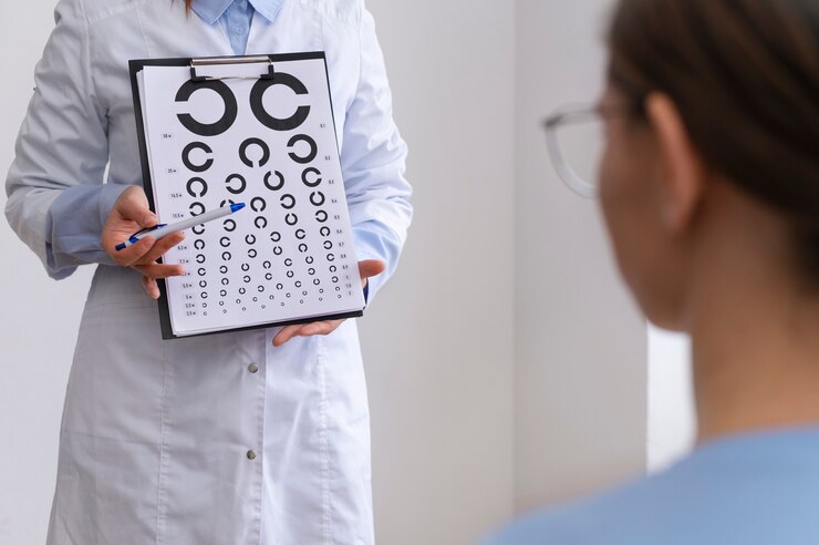
A Visual Acuity Test measures how well you can see at various distances. During this test, you will be asked to read letters on a chart (commonly known as a Snellen chart) from a specified distance. Each eye is tested separately, with and without corrective lenses if you wear them. The test determines the smallest letters you can read on the chart, indicating the clarity and sharpness of your vision. A decline in visual acuity may signal an underlying issue, such as retinal detachment, especially if it occurs suddenly.

Dilated Eye Exam

A dilated eye exam involves the use of special eyedrops to widen (dilate) the pupils, allowing the doctor to get a better view of the retina and other structures at the back of the eye. After administering the drops, which take about 15 to 30 minutes to fully dilate the pupils, the ophthalmologist uses a magnifying lens to inspect the retina for any tears, holes, or areas of detachment. The dilation provides a more comprehensive view, making it easier to detect abnormalities that might not be visible with non-dilated pupils. This examination can also reveal other eye conditions, such as macular degeneration or diabetic retinopathy.

Ophthalmoscopy

Ophthalmoscopy is a diagnostic procedure utilized to inspect the rear portion of the eye, encompassing the retina, optic disc, and blood vessels. The doctor uses an ophthalmoscope, which is a handheld instrument equipped with a light and several lenses. The ophthalmoscope allows for a detailed examination of the retina. The procedure can be performed directly, with the doctor looking through the pupil, or indirectly, using a special lens held close to the eye. Indirect ophthalmoscopy, often performed with the aid of scleral depression (pressing on the sclera or white of the eye), provides a wider view of the retina, which is especially useful for detecting peripheral retinal tears or detachments.

Ultrasound Imaging

Ultrasound imaging of the eye, also known as ocular ultrasonography, is used when retinal detachment is difficult to visualize due to opacities like vitreous Hemorrhage (bleeding into the vitreous). This non-invasive test involves placing a small probe on the closed eyelid after applying a gel to facilitate sound wave transmission. The probe emits high-frequency sound waves that bounce off the internal structures of the eye and create detailed images of the retina and surrounding tissues. These images can help the ophthalmologist identify the location and extent of a retinal detachment, as well as other possible abnormalities such as tumours or foreign bodies within the eye. Ultrasound is particularly useful in emergency settings where a clear view of the retina is obstructed.

TREATMENT

Laser Surgery (Photocoagulation): A laser is used to seal the retinal tear by creating small burns around it, preventing fluid from passing through.

Cryopexy: Freezing is used to reattach the retina by creating a scar that helps secure the retina to the eye wall.

Pneumatic Retinopexy: A gas bubble is injected into the vitreous cavity to push the retina back into place.

Scleral Buckling: A piece of silicone material is sewn onto the sclera (white of the eye) to push the wall of the eye against the detached retina.

Vitrectomy: Removal of the vitreous gel to relieve traction on the retina and replace it with a gas bubble or silicone oil.

A specialized hospital like Dr. Rani Menon Maxivision Eye Hospitals in Thrissur provides hope and enhance the quality of life for those affected by retinal detachment. Schedule a consultation with our experts. Our compassionate team at Dr. Rani Menon Maxivision Eye Hospitals is here to assist with all your diabetes-related health concerns, tailored to the type of diabetes you have.

Tags :
Blog
Share This :
Categories
Blog

The Importance of Regular Eye Exams

The Importance of Regular Eye Exams

eye-exam-clinic

The eyes are one of the most vital organs in the human body, playing a crucial role in our daily lives and overall well-being. Vision is often considered the most dominant of the senses, providing us with the ability to perceive and interpret the world around us. The importance of eyes can be understood through various facets of human experience, including health, safety, communication, and quality of life. The eyes are indispensable to human life, affecting nearly every aspect of our existence. Protecting our vision through regular eye check-ups, proper nutrition, and eye safety practices is essential for maintaining the richness and fullness of life that our eyes provide.

Health and Safety:

Our eyes serve as primary tools for navigating our environment safely. They help us detect hazards, recognize faces, and interpret signs, all of which are essential for avoiding accidents and staying safe. Good vision is critical for tasks that require precision and coordination, such as driving, reading, and operating machinery. Without a clear vision, these activities become challenging and potentially dangerous.

Communication:

Eyes are fundamental in non-verbal communication, allowing us to express and perceive emotions. Eye contact can convey confidence, interest, and understanding, playing a significant role in social interactions. Through our eyes, we can read the emotional states of others, fostering empathy and deeper connections.

Quality of Life:

The ability to see and appreciate the beauty of the world significantly enhances our quality of life. From enjoying a colorful sunset to appreciating art, vision enriches our experiences and adds depth to our perceptions. Visual stimuli are crucial for learning and cognitive development, especially in children, where visual aids are often used in education to facilitate better understanding.

Mental and Emotional Well-being:

Vision impairment can lead to social isolation and decreased independence, impacting mental health. People with good vision are more likely to engage in social activities and lead active lifestyles, contributing to their overall happiness and well-being.

Medical Insight:

Eyes can also be indicators of general health. Conditions like diabetes, hypertension, and even certain neurological disorders can manifest in the eyes, allowing for early diagnosis and treatment. Regular eye examinations can thus be pivotal in maintaining not just ocular health but overall health.


THE IMPORTANCE OF REGULAR EYE EXAMS

Regular eye exams are a critical component of maintaining overall health and well-being. Eyes are not only the windows to the world but also windows to our general health. Regular eye check-ups can detect eye problems at their earliest stages, when they are most treatable, and can also uncover other health issues.

Early Detection of Eye Diseases

One of the primary reasons for regular eye exams is the early detection of eye diseases. Many serious eye conditions, such as glaucoma, macular degeneration, and diabetic retinopathy, often develop without noticeable symptoms in the early stages. By the time symptoms become apparent, significant, irreversible damage may have already occurred. Regular eye exams can identify these diseases early, allowing for timely intervention that can prevent or minimize vision loss.

Monitoring Vision Changes

Vision changes can occur gradually and might go unnoticed until they significantly impact daily activities. Regular eye exams can detect subtle changes in vision that you might not be aware of. For example, changes in refractive errors such as myopia (near sightedness), hyperopia (far sightedness), and astigmatism can be easily corrected with updated prescriptions for glasses or contact lenses. Keeping your prescription current ensures that you can see as clearly and comfortably as possible.

Identifying General Health Issues

The eyes can reveal a lot about overall health. Conditions such as diabetes, hypertension, high cholesterol, and even certain cancers can manifest in the eyes. Through a comprehensive eye exam, an optometrist or ophthalmologist can spot signs of these systemic diseases and refer you to appropriate healthcare professionals for further evaluation and treatment. For instance, diabetic retinopathy is a common complication of diabetes that can be detected through an eye exam, often before the patient is aware of their diabetic condition.

Children’s Eye Health

Regular eye exams are particularly important for children. Vision is essential for a child’s development and learning.  Undetected vision problems can lead to difficulties in school and can impact a child’s ability to reach developmental milestones. Early eye exams can catch issues such as amblyopia (lazy eye), strabismus (crossed eyes), and other refractive errors, ensuring that children have the visual skills they need to succeed academically and socially.

Age-Related Eye Conditions

As we grow older, the likelihood of developing eye conditions such as cataracts, glaucoma, and macular degeneration rises. Regular eye exams become increasingly important for older adults to monitor for these conditions. Early detection and management can help preserve vision and maintain quality of life.

Eye Safety and Preventive Care

Regular eye exams also provide an opportunity to discuss eye safety and preventive care. Eye care professionals can offer advice on protecting your eyes from injury, reducing eyestrain, and maintaining eye health through proper nutrition and lifestyle choices. For instance, they can recommend wearing protective eyewear during certain activities or using appropriate lighting and ergonomic setups to minimize eyestrain.

Conclusion

Regular eye exams are essential for maintaining good vision and overall health. They enable the early detection and treatment of eye diseases, monitor vision changes, and can uncover systemic health issues. For children, they are vital in ensuring proper visual development, and for older adults, they help manage age-related eye conditions. Additionally, regular exams offer a chance to receive valuable advice on eye safety and preventive care. Prioritizing kids is a simple yet powerful way to protect one of your most important senses and maintain your overall well-being.


If you are considering an eye check-up for yourself or for your loved ones, consult a professional eye specialist only. Once you have decided to proceed, schedule an initial consultation

Tags :
Blog
Share This :