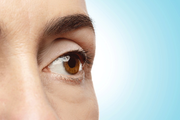
Contact Number +91 98468 78222
Mon-Sat: 8AM – 7PM

Categories
Blog

Detecting Glaucoma: The Importance of Regular Eye Exams

Detecting Glaucoma: The Importance of Regular Eye Exams

Recent Posts

Have Any Questions?

Please contact us, if you have any queries

Categories

What is glaucoma?

     Glaucoma is a group of eye diseases that damage the optic nerve, which carries visual information from the eye to the brain. Glaucoma is often caused by increased eye pressure, but it can also occur in people with normal eye pressure.

Why is it important to detect glaucoma early?

     Glaucoma is a leading cause of blindness in adults, but it is often preventable with early detection and treatment. Glaucoma usually progresses without any symptoms, so it is important to have regular eye exams, even if you have good vision.

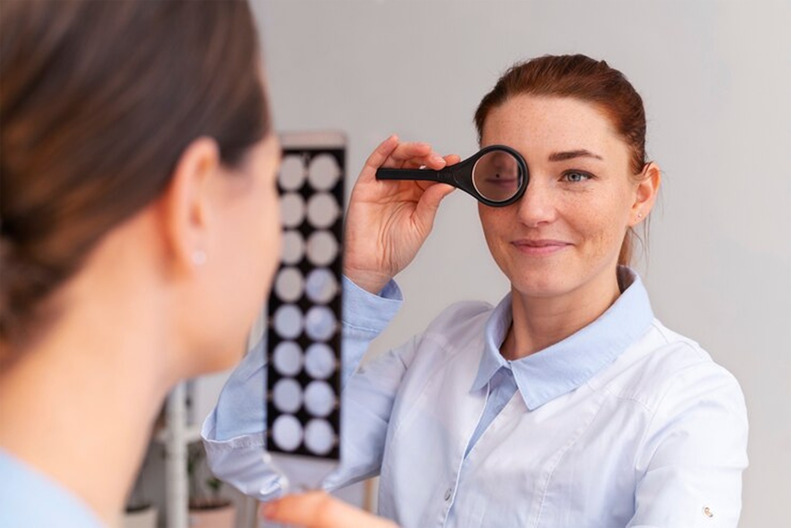
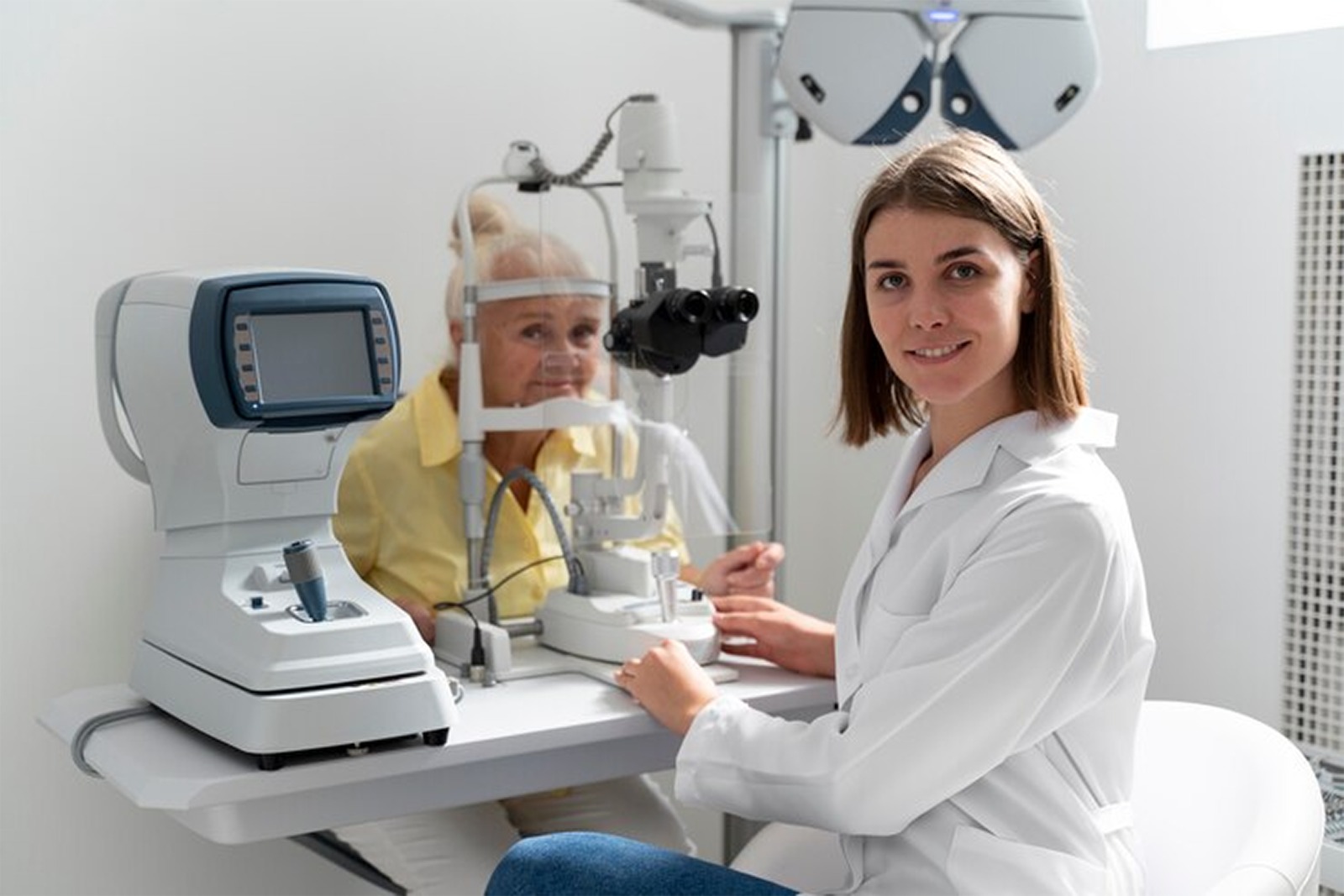
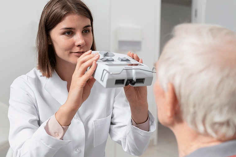
How is glaucoma detected?

     A comprehensive eye exam includes a variety of tests to check for glaucoma, including:

  • Tonometry: This test measures the pressure inside the eye.
  • Ophthalmoscopy: The doctor will examine the optic nerve and retina for signs of damage.
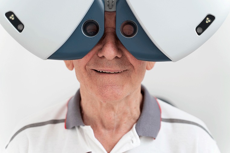
  • Visual field test: This test measures the range of peripheral vision.

Who is at risk for glaucoma?     

     Certain people are at higher risk for developing glaucoma, including:

  • People over the age of 40
  • People with a family history of glaucoma
  • People with high eye pressure
  • People with diabetes
  • People with a history of eye injuries

How is glaucoma treated?

     There is no cure for glaucoma, but there are treatments to slow or stop the progression of the disease and prevent vision loss. Treatment options include eye drops, laser surgery, and conventional surgery.

The importance of regular eye exams

     Regular eye exams are the best way to detect glaucoma early and prevent vision loss. The American Academy of Ophthalmology recommends that adults have a comprehensive eye exam every two years, or more often if they are at high risk for glaucoma.

Here are some tips for getting the most out of your eye exam:

  • Be sure to tell your eye doctor about any medical conditions you have or medications you are taking.
  • Bring a list of all your medications to your appointment.
  • Be honest with your eye doctor about your vision and any concerns you have.
  • Ask your eye doctor about your risk for glaucoma and how often you should have your eyes examined.

     Glaucoma is a serious eye disease, but it is often preventable with early detection and treatment. Regular eye exams, such as those conducted at Dr. Rani Menon’s Eye Clinic in Trissur, are the best way to detect glaucoma early and protect your vision. Dr. Rani Menon’s Eye Clinic is dedicated to providing comprehensive eye care services, ensuring that patients receive the necessary screenings and treatment options to maintain their eye health. So, don’t wait – schedule your regular eye exam today to safeguard your vision and prevent the onset of glaucoma.

Tags :
Blog
Share This :
Categories
Blog

Retinal Damage in Myopia: Risks and Strategies for High Myopia Patients

Retinal Damage in Myopia: Risks and Strategies for High Myopia Patients

     Myopia, also known as near-sightedness, is a common eye condition in which people can see close objects clearly but distant objects appear blurry. Myopia occurs when the eyeball is too long or the cornea (the clear front part of the eye) is too curved, causing light to focus in front of the retina instead of on it.

     Myopia is a progressive condition, meaning that it can worsen over time. High myopia, defined as having a prescription of -6.00 diopters (D) or higher, is associated with an increased risk of retinal damage.

How does myopia cause retinal damage?

     The retina is a light-sensitive tissue that lines the back of the eye. It contains millions of photoreceptor cells that convert light into electrical signals that are sent to the brain. The brain then interprets these signals to create images.

     In high myopia, the elongation of the eyeball can cause the retina to stretch and become thinner. This can lead to a number of retinal problems, including:

  • Retinal detachment: This is a serious condition in which the retina separates from the choroid, the layer of blood vessels that nourishes the retina. Retinal detachment can cause permanent vision loss.
  • Myopic macular degeneration: This is a condition that damages the macula, the central part of the retina responsible for sharp central vision. Myopic macular degeneration can lead to vision loss and blindness.
  • Glaucoma: This is a group of eye diseases that damage the optic nerve. Glaucoma can lead to vision loss and blindness.

Strategies for high myopia patients

     There are a number of things that high myopia patients can do to reduce their risk of retinal damage, including:

  • Get regular eye exams: It is important for high myopia patients to have regular eye exams to monitor for signs of retinal damage. Eye exams should include dilation of the pupils, which allows the doctor to get a better view of the retina.
  • Use corrective lenses: Wearing corrective lenses, such as glasses or contact lenses, can help to focus light properly on the retina.
  • Avoid smoking: Smoking can increase the risk of retinal detachment and other eye problems.
  • Eat a healthy diet: Eating a healthy diet that includes plenty of fruits, vegetables, and whole grains can help to protect the retina.
  • Manage underlying medical conditions: High myopia patients should also manage any underlying medical conditions, such as diabetes and hypertension.


New treatments for retinal damage in high myopia patients

     There are a number of new treatments emerging for retinal damage in high myopia patients. These treatments include:

  • Anti-VEGF injections: These injections can help to stop the growth of new blood vessels in the retina, which can lead to retinal detachment and other problems.
  • Laser surgery: Laser surgery can be used to create small scars in the retina, which can help to strengthen it and reduce the risk of retinal detachment.
  • Implantable devices: Implantable devices can be used to support the retina and reduce the risk of retinal detachment.

     If you are a high myopia patient, talk to your doctor about the best way to manage your condition and reduce your risk of retinal damage.

     Dr. Rani Memon’s Eye Clinic in Trissur is a leading eye care center in India that specializes in the treatment of high myopia and retinal damage. The clinic offers a wide range of services, including comprehensive eye exams, laser surgery, and implantable devices.

     If you are a high myopia patient and are looking for expert care, contact Dr. Rani Memon’s Eye Clinic in Trissur.


Tags :
Blog
Share This :
Categories
Blog

Understanding Corneal Blindness: Who Needs a Cornea Transplant

Understanding Corneal Blindness: Who Needs a Cornea Transplant

     Corneal blindness is a condition in which the cornea, the clear front part of the eye, becomes damaged or scarred, causing vision loss. The cornea is responsible for focusing light onto the retina, the light-sensitive tissue at the back of the eye. When the cornea is damaged, light cannot focus properly, resulting in blurred vision or even blindness.

What are the causes of corneal blindness?

     There are many causes of corneal blindness, including:

  • Injury: The cornea can be injured by a sharp object, such as a finger, a pencil, or a piece of glass. Chemical burns and sports injuries can also damage the cornea.
  • Infection: Infections of the cornea, such as keratitis, can cause scarring and vision loss.
  • Diseases: Certain eye diseases, such as keratoconus, Fuchs dystrophy, and bullous keratopathy, can damage the cornea and lead to blindness.
  • Other causes: Other causes of corneal blindness include malnutrition, vitamin A deficiency, and congenital defects.

Who needs a cornea transplant?

     A cornea transplant is a surgical procedure in which the damaged or scarred cornea is replaced with a healthy cornea from a deceased donor. Cornea transplants are performed to restore vision and relieve pain or other symptoms associated with corneal blindness.

A cornea transplant may be recommended for people with corneal blindness caused by:

  • Injury
  • Infection
  • Disease
  • Complications from previous eye surgery
  • Genetic condition

How is a cornea transplant performed?

     A cornea transplant is a relatively safe and effective procedure. The surgery is usually performed under general anesthesia. During the surgery, the surgeon will remove the damaged or scarred cornea and replace it with a healthy donor cornea.

There are two main types of cornea transplants:

  • Penetrating keratoplasty (PKP): This is the most common type of cornea transplant. In PKP, the entire cornea is replaced.
  • Endothelial keratoplasty (EK): This type of cornea transplant replaces only the inner layer of the cornea. EK is less invasive than PKP and has a shorter recovery time.

What are the risks and benefits of a cornea transplant?

     The risks of a cornea transplant include:

  • Infection
  • Bleeding
  • Cataracts
  • Glaucoma
  • Rejection of the donor cornea

     The benefits of a cornea transplant usually outweigh the risks. Cornea transplants can restore vision and improve quality of life for people with corneal blindness.

How to prepare for a cornea transplant

     If you are scheduled for a cornea transplant, your doctor will give you specific instructions on how to prepare for surgery. These instructions may include:

  • Stopping certain medications
  • Having someone drive you to and from surgery


What to expect after a cornea transplant

     After a cornea transplant, you will need to rest and avoid strenuous activity. You will also need to take eye drops and medications to prevent infection and inflammation.

     Most people recover from a cornea transplant within a few months. However, it is important to follow your doctor’s instructions carefully and to attend all follow-up appointments.

     Cornea transplants are a safe and effective way to restore vision for people with corneal blindness. If you are considering a cornea transplant, it is essential to have a thorough discussion with your doctor about the risks and benefits of the procedure. For those seeking expert care and guidance in cornea transplantation, consider reaching out to Dr. Rani Memon’s Eye Clinic in Trissur. Their dedicated team specializes in comprehensive eye care services and can provide valuable insights into your treatment options. Restoring your vision and improving your quality of life is their top priority, and they are ready to assist you on your journey to better eye health.


Tags :
Blog
Share This :
Categories
Blog

Surgical solutions for Glaucoma: When is surgery necessary?

Surgical solutions for Glaucoma: When is surgery necessary?

     Glaucoma is a group of eye diseases that damage the optic nerve, which carries visual information from the eye to the brain. Glaucoma is often caused by increased eye pressure, but it can also occur in people with normal eye pressure.

     Glaucoma is a leading cause of blindness in adults, but it is often preventable with early detection and treatment. However, in some cases, surgery may be necessary to lower eye pressure and prevent vision loss.

When is surgery necessary for glaucoma?

Surgery may be recommended for glaucoma if:

  • Eye drops or laser surgery have not been effective in lowering eye pressure.
  • Eye pressure is very high and is causing damage to the optic nerve.
  • Glaucoma is causing vision loss.
  • Glaucoma is associated with other eye problems, such as cataracts.

Types of glaucoma surgery

There are two main types of glaucoma surgery:

  • Trabeculectomy: This is the most common type of glaucoma surgery. It involves creating a new drainage channel in the eye to allow fluid to escape more easily.
  • Tube shunt surgery: This type of surgery involves placing a small tube in the eye to drain fluid into another part of the eye or to the surface of the eye.

Minimally invasive glaucoma surgery (MIGS)

     MIGS is a newer group of glaucoma surgeries that are less invasive and have shorter recovery times than traditional glaucoma surgeries. MIGS procedures are often combined with cataract surgery.

Risks and benefits of glaucoma surgery

Glaucoma surgery is a safe and effective procedure, but there are some risks, such as:

  • Infection
  • Bleeding
  • Cataracts
  • Scarring
  • Low eye pressure

     The benefits of glaucoma surgery usually outweigh the risks. Glaucoma surgery can lower eye pressure and prevent vision loss.

How to prepare for glaucoma surgery

     Your doctor will give you specific instructions on how to prepare for glaucoma surgery. These instructions may include:

  • Stopping certain medications
  • Having someone drive you to and from surgery

What to expect after glaucoma surgery

After glaucoma surgery, you will need to rest and avoid strenuous activity. You will also need to take eye drops and medications to prevent infection and inflammation.

Most people recover from glaucoma surgery within a few weeks. However, it is important to follow your doctor’s instructions carefully and to attend all follow-up appointments.

Here are some additional tips for people considering glaucoma surgery:

  • Ask your doctor about all of your treatment options and the risks and benefits of each.
  • Make sure you have a good support system in place to help you recover from surgery.
  • Be patient and follow your doctor’s instructions carefully.

    In conclusion, glaucoma surgery is a safe and effective way to lower eye pressure and prevent vision loss. If you have glaucoma, talk to your doctor about whether surgery is right for you. For expert care and guidance in managing your glaucoma, consider reaching out to Dr. Rani Menon’s Eye Clinic in Trissur. Their experienced team specializes in comprehensive eye care services and can help you make informed decisions regarding your treatment, whether it involves surgery or other therapeutic options. Remember that early intervention and professional guidance are key to protecting your vision and preventing the progression of glaucoma.

Tags :
Blog
Share This :
google-site-verification=toJuA_RRy2ANvvRwUeAvEpKhQZGVqSkYAllfXhlKqtY