Cataract Surgery in Thrissur: Procedure, Cost & Recovery (2026 Guide)
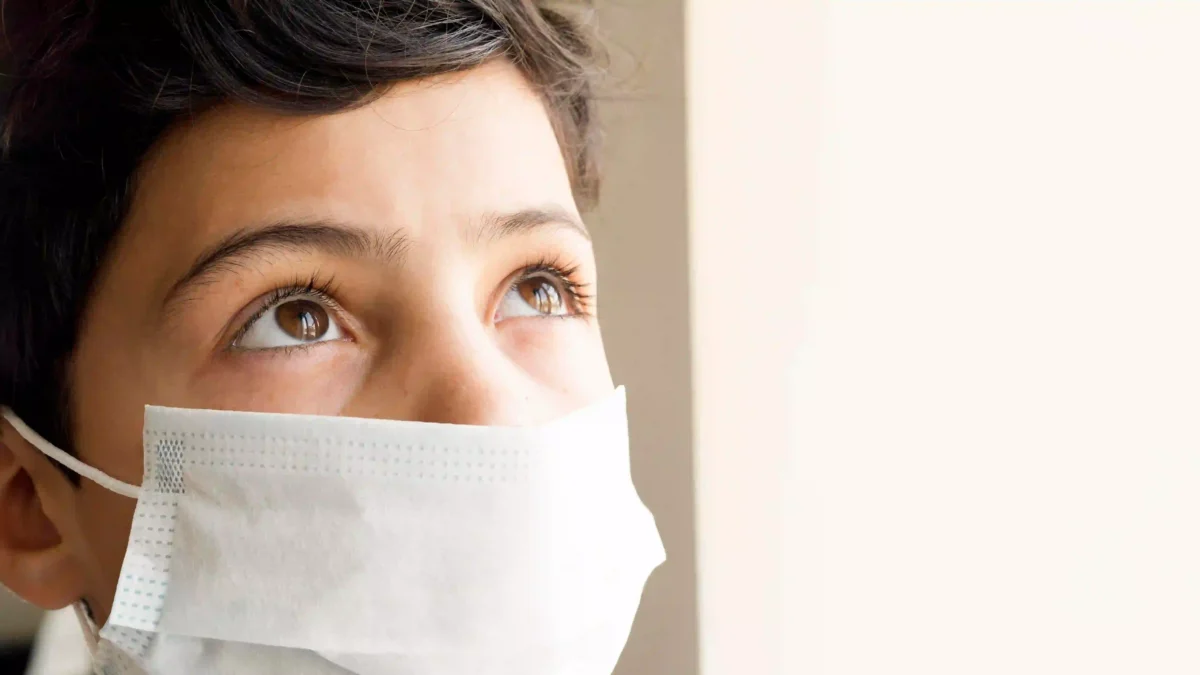
Sight is something most of us take for granted — until it starts to fade. If the world around you has begun to look washed out, blurred, or wrapped in a hazy glow, you might be experiencing the early stages of a cataract. It is one of the most common eye conditions in India, and thankfully, one of the most treatable.
Thrissur, often called the cultural capital of Kerala, has quietly grown into a hub for advanced eye care. Patients from across the state — and sometimes from neighbouring Tamil Nadu and Karnataka — travel here for cataract treatment. The combination of experienced ophthalmologists, modern surgical technology, and relatively accessible costs makes Thrissur a practical choice for many families.
This guide covers everything you need to know before you or a loved one walks into an eye hospital in Thrissur: what the surgery involves, what it costs, how recovery unfolds, and what questions to ask your doctor.
What Exactly Is Cataract Surgery?
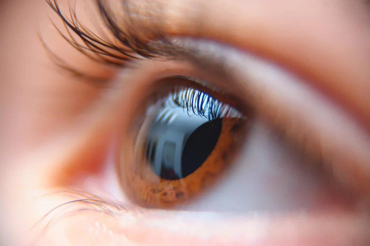
The lens of your eye is naturally clear. Over time — usually due to ageing, but sometimes because of diabetes, prolonged steroid use, or UV exposure — proteins inside the lens begin to clump together and turn cloudy. This clouding is a cataract.
Cataract surgery removes that clouded lens and replaces it with a small, artificial lens called an intraocular lens (IOL). The procedure does not restore perfect vision on its own; it restores optical clarity. Depending on the type of IOL chosen, many patients find they need glasses far less — or not at all — after surgery.
It is one of the most performed elective surgeries worldwide, with a safety record that has improved dramatically over the past decade. In 2026, the surgical techniques available in Thrissur are on par with what you would find in major metropolitan centres.
Signs You May Need Cataract Surgery
Cataracts develop slowly, which means many people adapt to worsening vision without realising just how much they have lost. Here are signs that it may be time to see a cataract specialist in Thrissur:
- Blurred or cloudy vision that glasses cannot fully correct
- Increased sensitivity to bright lights, particularly headlights while driving at night
- Colours appearing dull, yellowish, or faded
- Frequent changes in your eyeglass or contact lens prescription
- Double vision in one eye
- Difficulty reading fine print even with reading glasses
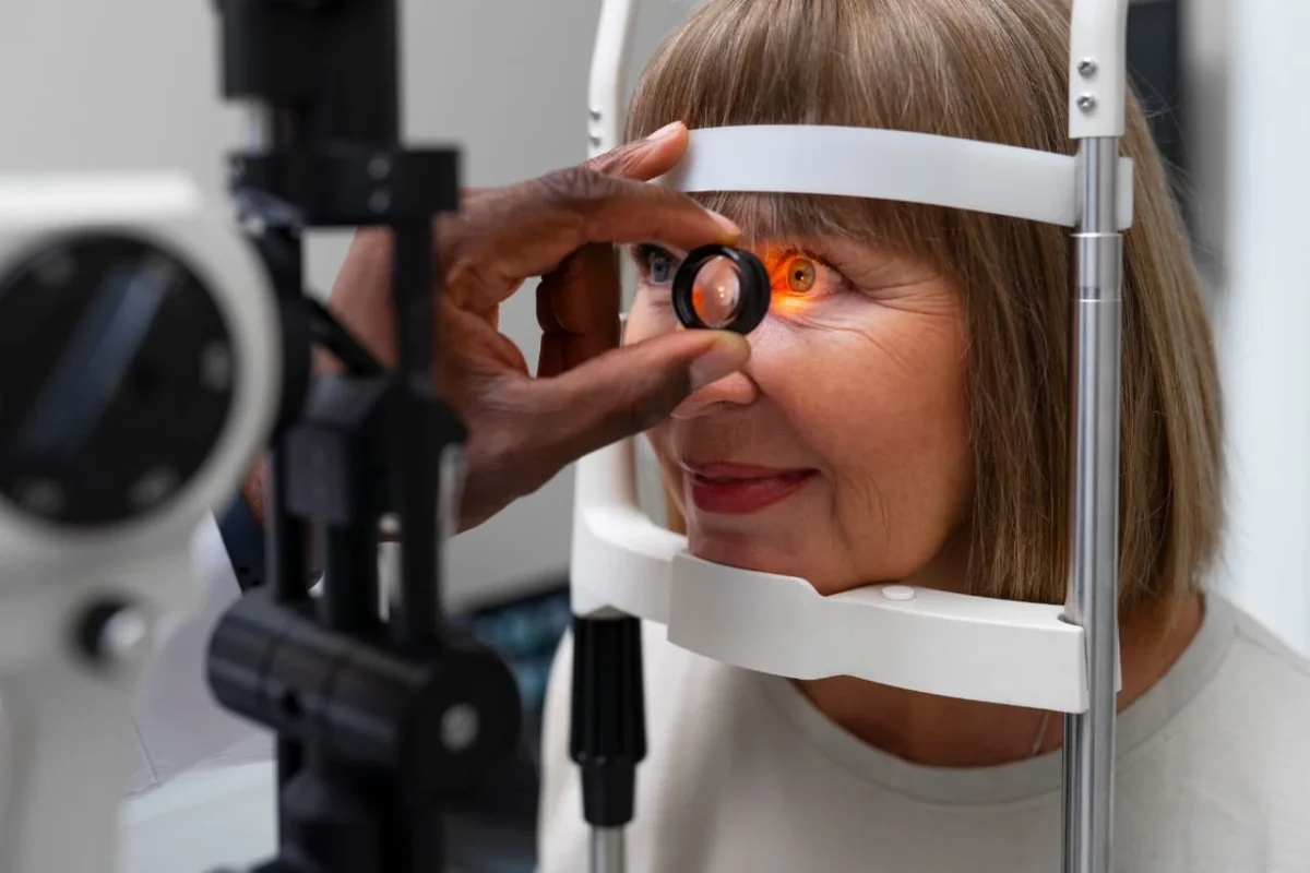
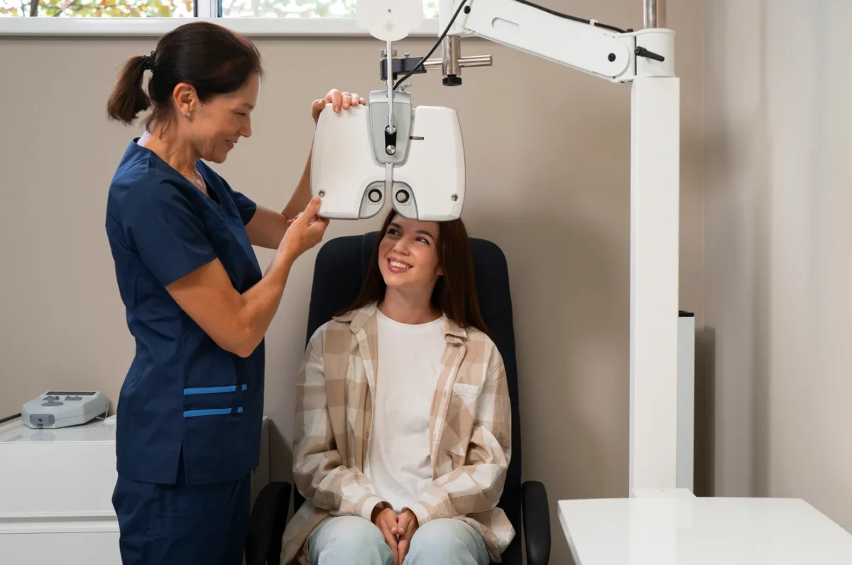
If two or more of these sound familiar, an ophthalmologist can confirm the diagnosis with a dilated eye exam. Surgery is typically recommended when cataracts begin to interfere with daily activities — not just because they are present.
Types of Cataract Surgery Available in Thrissur
Eye hospitals in Thrissur offer several surgical approaches, each suited to different patient needs and budgets.
Phacoemulsification (Phaco Surgery)
This is the gold standard for cataract removal and the most widely performed technique across Thrissur. A tiny ultrasonic probe is inserted through a self-sealing incision of roughly 2–3 mm. The probe breaks the clouded lens into microscopic fragments using sound waves, which are then gently suctioned out. A foldable IOL is placed through the same incision.
Phaco cataract surgery in Thrissur is popular for good reason: it requires no stitches, causes minimal trauma to surrounding tissue, and most patients recover usable vision within 24 to 48 hours.
Laser-Assisted Cataract Surgery (LACS / Femto-Laser)
Laser cataract surgery in Thrissur is available at select advanced eye centres. A femtosecond laser — the same technology used in LASIK — performs the most precise steps of the procedure: making the corneal incision, opening the lens capsule, and softening the cataract before the surgeon removes it.
The laser reduces the amount of ultrasonic energy used during phaco, which may translate to a faster visual recovery and a lower risk of corneal swelling. It is particularly useful for patients with dense cataracts or pre-existing corneal conditions. The trade-off is cost — laser-assisted surgery carries a higher price tag than standard phaco.
Lens Replacement Options
The artificial lens implanted during surgery shapes your visual outcome. Choices include:
- Monofocal IOL — corrects vision at one distance (usually far). Reading glasses are still needed.
- Multifocal IOL — multiple focal points; reduces or eliminates dependence on glasses for both near and distance.
- Toric IOL — designed specifically for patients with astigmatism.
- Extended Depth of Focus (EDOF) IOL — provides a continuous range of vision with fewer halos than multifocal lenses.
Your surgeon will recommend a lens based on your prescription, lifestyle, and the health of your cornea. There is no single “best” IOL — it depends on what matters most to you.
Step-by-Step: What Happens During Cataract Surgery
Understanding the procedure often eases anxiety. Here is what typically happens on the day of surgery at an eye hospital in Thrissur:
- Pre-operative preparation — Dilating eye drops are administered 30–60 minutes before surgery. Your blood pressure and vitals are checked.
- Anaesthesia — Local anaesthetic drops are applied to numb the eye surface. Most patients are awake throughout; general anaesthesia is rarely needed and usually reserved for children or patients with special needs.
- Incision — A microincision is made at the edge of the cornea.
- Capsulotomy — A circular opening is made in the front of the lens capsule to access the cataract.
- Phacoemulsification — The clouded lens is broken up and suctioned out.
- IOL Implantation — The chosen artificial lens is inserted in a folded state through the same incision. It unfolds and settles into position.
- Closure — The incision is self-sealing in most cases. No stitches are placed. An eye shield is taped over the eye.
How Long Does Cataract Surgery Take?
The actual surgical time for phacoemulsification is typically 10 to 20 minutes per eye. However, account for the full hospital visit — pre-operative preparation, waiting, and the brief post-operative observation period — which adds up to 2 to 4 hours.
Both eyes are almost never operated on the same day. Surgeons typically wait 1 to 4 weeks between procedures to ensure the first eye heals well before treating the second.
Good to know: Most patients are discharged the same day with a protective eye shield and a bag of prescribed drops. You will need someone to drive you home. |
Recovery Timeline After Cataract Surgery
Recovery from cataract surgery is generally smooth, but it is not instant. Here is what most patients experience:
- Day 1: Vision is often blurry and the eye may feel gritty or mildly sore. This is normal. Rest with the eye shield in place.
- Days 2–3: Many patients notice a significant improvement in clarity. Colours may appear brighter than expected — almost vivid — because the clouded lens has been removed.
- Week 1: Light activity is fine. Avoid rubbing the eye. Attend your first follow-up appointment.
- Weeks 2–4: Most patients return to normal daily activities including reading, cooking, and light exercise. Driving clearance depends on your surgeon’s assessment.
- 4–6 weeks: The eye stabilises. A final spectacle prescription, if needed, is given at this point.
Full healing of the internal structures takes approximately 8 weeks. Some patients notice mild visual fluctuations during this period — this is normal and settles on its own.
On recovery time: Cataract recovery time is short compared to most surgeries. The majority of patients are back to light work within a week, though individual timelines vary based on the type of IOL and any co-existing eye conditions. |
Dos and Don’ts After Cataract Surgery
What to do
- Use all prescribed eye drops exactly as directed — typically antibiotic and anti-inflammatory drops for 3–4 weeks
- Wear the eye shield at night for the first two weeks to prevent accidental rubbing during sleep
- Attend all scheduled follow-up appointments
- Wear sunglasses outdoors to protect the healing eye from UV and dust
What to avoid
- Rubbing or pressing the eye, even gently
- Swimming, hot tubs, or submerging the face in water for at least 4 weeks
- Heavy lifting or strenuous exercise for 2–4 weeks
- Dusty or smoky environments without eye protection
- Skipping eye drops even if vision seems fine
Possible Risks and Side Effects
Cataract surgery is considered very safe, with a complication rate below 2% in experienced hands. That said, all surgeries carry some risk. The more common, temporary side effects include:
- Mild redness, watering, or a foreign body sensation for the first few days
- Sensitivity to bright light
- Seeing halos or starbursts around lights, especially at night (more common with multifocal lenses)
Less common but more serious complications — such as infection, bleeding, retinal detachment, or posterior capsule opacification (a secondary clouding that can develop months to years later and is easily treated with a brief laser procedure) — should be discussed with your surgeon before the operation.
The risk of serious complications is significantly lower when surgery is performed at an accredited facility by an experienced ophthalmologist. This is one reason choosing the right hospital matters.
How to Choose the Best Eye Hospital in Thrissur
Not all eye hospitals are equal. When evaluating your options for cataract treatment in Thrissur or anywhere in Kerala, consider:
- Surgeon experience — How many cataract surgeries does the surgeon perform annually? Experienced surgeons have lower complication rates.
- Technology — Does the hospital offer phaco and femtosecond laser options? Are OCT and biometry measurements taken pre-operatively for accurate IOL power calculation?
- IOL range — A hospital that offers a wide selection of IOLs (monofocal, toric, multifocal, EDOF) allows you to choose based on your visual goals, not just what is available.
- Post-operative care — Is there a dedicated follow-up protocol? Can you reach someone quickly if a concern arises after surgery?
- Transparency on costs — Reputable centres provide a clear breakdown of charges before you commit, with no surprise fees.
- Accreditation — NABH accreditation or equivalent is a reliable indicator of quality standards.
It is perfectly reasonable to consult at more than one facility before deciding. A trustworthy hospital will welcome your questions rather than rush you into surgery.
Why Patients Across Kerala Seek Advanced Cataract Treatment in Thrissur
Thrissur has a concentration of eye care expertise that is relatively rare outside the major metros. Several well-established ophthalmology centres in the city have invested in technology — femtosecond laser platforms, advanced IOL calculation systems, and intraoperative aberrometry — that was once available only in Chennai or Kochi.
The city’s geography helps too. Thrissur is well connected by road and rail to Palakkad, Malappuram, Ernakulam, and parts of northern Kerala. For patients in smaller towns without access to sub-specialty eye care, making the journey to Thrissur for a procedure is genuinely practical.
Word of mouth also plays a significant role. Families that have had good outcomes tend to recommend the same surgeon or hospital to relatives. Over time, this has built a reputation for cataract treatment in Kerala that extends well beyond the city limits.
Frequently Asked Questions
Is cataract surgery painful?
No. Anaesthetic drops numb the eye surface completely before surgery. You may feel mild pressure during the procedure but not pain. Any discomfort afterwards is usually manageable with standard pain relief and settles within a day or two.
At what age should cataracts be operated on?
There is no fixed age. Surgery is recommended when the cataract affects your quality of life or daily function — not simply because it is present. Some patients are operated on in their 50s if the cataract is dense; others wait until their 80s. Your ophthalmologist will advise based on your specific situation.
How long do the results of cataract surgery last?
The artificial lens implanted during surgery is designed to last a lifetime. Cataracts do not recur in the treated eye. However, a condition called posterior capsule opacification (PCO) can cause cloudy vision months or years after surgery. This is easily resolved with a quick, painless laser procedure called YAG capsulotomy.
Can I have cataract surgery if I have diabetes?
Yes, in most cases. Diabetic patients can safely undergo cataract surgery, though blood sugar levels should be well controlled beforehand. Your ophthalmologist may also assess for diabetic retinopathy, which can affect the visual outcome after surgery.
What is the difference between phaco and laser cataract surgery?
In phacoemulsification (phaco), the surgeon manually performs all steps of the procedure using a handheld probe. In laser-assisted cataract surgery, a femtosecond laser handles the most precise steps — the incision, capsulotomy, and lens softening — before the surgeon completes the removal. Laser surgery offers marginally greater precision, especially for astigmatism correction, but both techniques have excellent safety records in experienced hands.
Will I still need glasses after cataract surgery?
That depends on the IOL you choose. With a standard monofocal lens, most patients see well at distance but still need reading glasses. Multifocal and EDOF lenses are designed to reduce this dependence significantly. Your surgeon will discuss realistic expectations based on your eye measurements during the pre-operative workup.
How soon can I travel after cataract surgery?
Short car journeys are usually fine within a day or two. Air travel is generally permitted after 1 week, though it is advisable to carry your eye drops in your hand luggage and stay well hydrated. Inform your surgeon if you have a long trip planned immediately after surgery.
Making the Decision
Cataract surgery is one of the most transformative procedures in medicine — simple in concept, profound in outcome. For many patients, the change is striking: colours look brighter, reading becomes easier, and driving at night is no longer something they dread.
If you are noticing the signs, the most useful thing you can do is book a consultation with a cataract specialist in Thrissur. A thorough examination takes less than an hour and gives you a clear picture of whether — and when — surgery makes sense for you. There is no obligation to proceed immediately.
Good vision is worth protecting. And in 2026, the tools and expertise needed for safe, effective cataract treatment in Thrissur are closer than many people realise.
Next step: Call your chosen eye hospital in Thrissur to schedule a cataract evaluation. Bring your current glasses and a list of any medications you take. The rest follows from there. |
This article is intended for general information only and does not constitute medical advice. Please consult a qualified ophthalmologist for diagnosis and treatment decisions.