Corneal abrasions, or scratches on the cornea is a common injury that can cause severe discomfort and fear, especially among children. The cornea, a transparent, dome-shaped surface that covers the front of the eye, is essential for vision and eye protection. This blog covers the cause, symptoms, diagnosis, and treatment of corneal abrasions, providing useful information for parents and guardians.
What is a Corneal Abrasion?
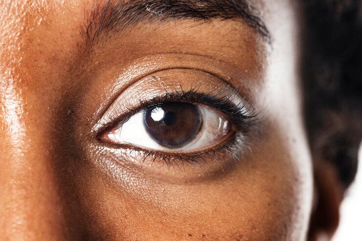
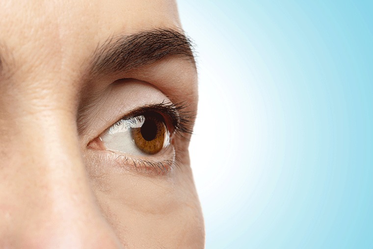
A corneal abrasion is a scratch or injury to the cornea, known as the transparent layer of the eye that protects the iris and pupil. Although the cornea is intended to be protective, it is readily injured by external elements or trauma. Corneal abrasions are especially common in children, due to their active play and curiosity.
Understanding the cornea and its role
The cornea is a key component of the eye’s structure, affecting both vision and eye protection. It acts as a barrier to dirt, bacteria, and other particles that might hurt the eyes. The cornea also helps to concentrate light, resulting in clearer vision. Because of its exposure, the cornea is vulnerable to injury, particularly in youngsters who are more prone to accidents at play.
Common Causes of Corneal Abrasions
Foreign Bodies: Dust, debris, or tiny particles can cause corneal scratches.
Trauma from Objects: Toys, fingernails and sharp objects could lead to eye trauma.
Contact Lenses: Improperly fitted or poorly maintained lenses might cause abrasion
Chemical Exposure: Chemical exposure can cause corneal injury.
Emphasizing the Need for Regular Eye Check-Ups
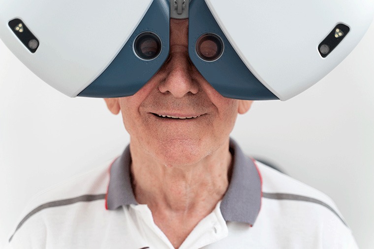
Routine eye exams are necessary for the early diagnosis of corneal abrasions and other eye problems. Parents should ensure that their children have frequent eye exams, especially if they engage in activities that raise the risk of eye injury. Early detection and treatment can prevent minor abrasions from progressing to more serious complications.
Symptoms of Corneal Abrasions
Children experiencing a corneal abrasion might have various kinds of symptoms. These symptoms may vary depending on the degree of the damage and the child’s response. Common signs include:
- Eye Pain:Common symptoms of the afflicted eye include sharp or agonizing discomfort.
- Redness:The eye can appear red and irritated.
- Tearing: Excessive watering of the eye is commonly observed.
- Light Sensitivity: Light sensitivity causes discomfort when exposed to bright lights.
- Blinking and Eye Rubbing:Persistent blinking or eye rubbing.
Symptoms in younger children may include difficulties opening the eye or an increased tendency to keep the eye closed.
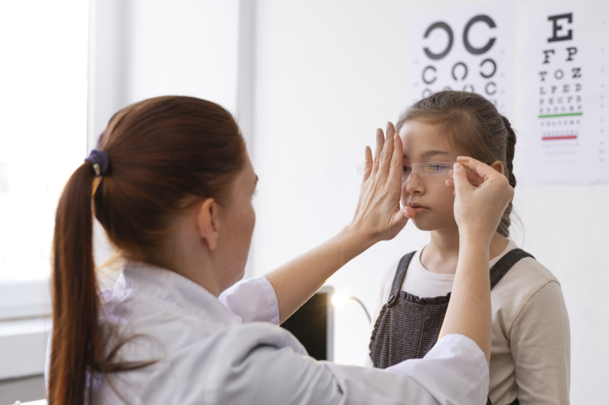
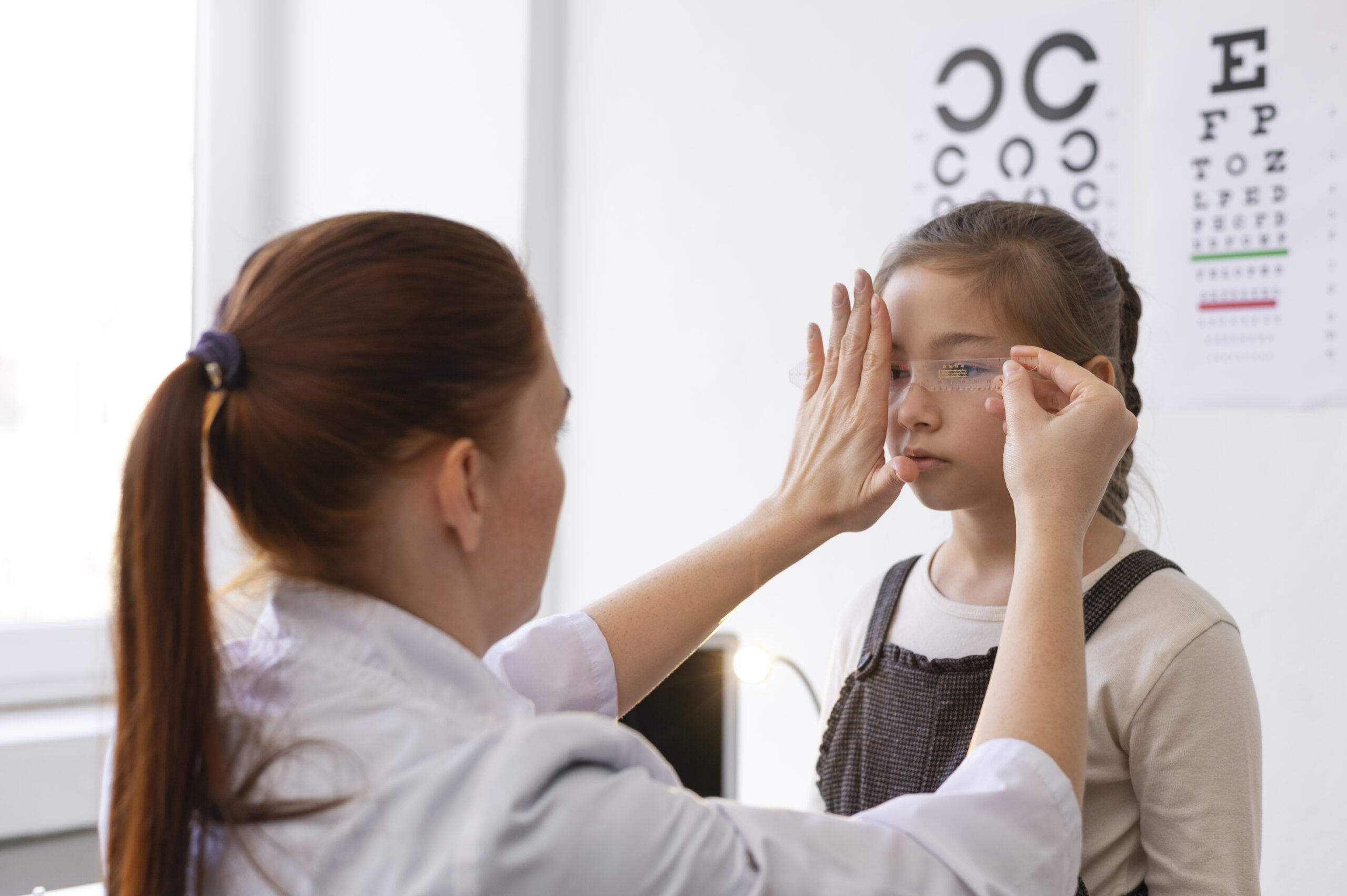
Diagnosing Corneal Abrasions
A comprehensive examination by an eye professional is necessary to diagnose corneal abrasions. The diagnostic process often includes:
Medical History: Examine the child’s symptoms, recent injuries, and exposure to foreign objects.
Physical Examination: The ophthalmologist will do a thorough eye examination, which may include:
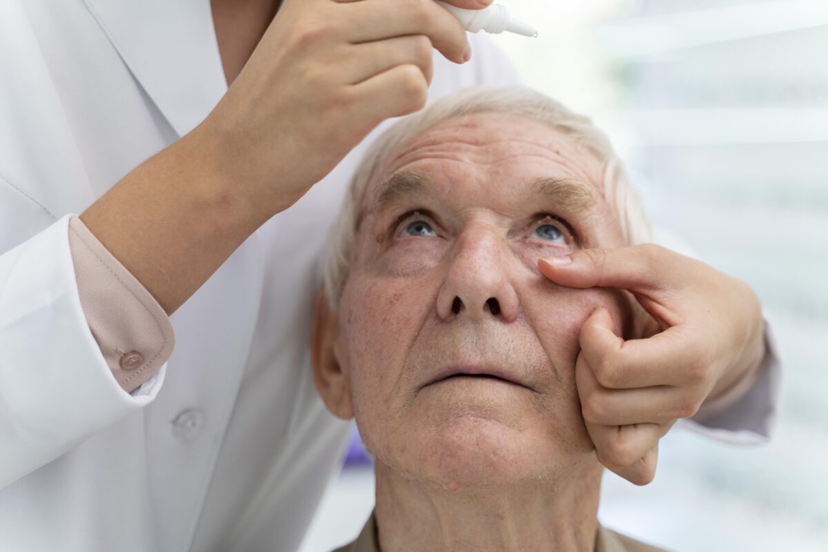
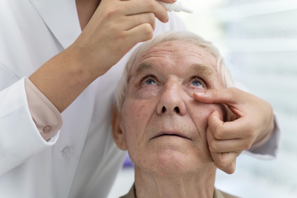
Numbing Drops: Local anesthetic drops are used to reduce pain during examinations.
Fluorescein Stain: Staining involves applying a yellow dye to the eye to emphasize abrasions under a blue light.
Treatment Options for Corneal Abrasions
Most corneal abrasions heal within a few days if properly treated. The primary objectives are to alleviate pain, avoid infection, and assure full recovery. Treatment may include:
- Removing Foreign Bodies: To remove a foreign item from the eye, use a cotton applicator or saline solution.
- Medication: Antibiotic ointments or drops may be recommended to prevent infection and aid healing.
- Eye Patch: Applying a patch helps relieve pain and protect the eye and patches are typically used for 12 to 24 hours.
- Pain Management: Over-the-counter pain medications may be used to alleviate discomfort.
A referral is required for severe abrasions or if problems emerge, such as a corneal ulcer.
Preventive Measures
Preventing corneal abrasions involves adopting measures to protect the eyes from harm.
Protective Eyewear: Wear protective goggles or glasses during risky activities like sports or outdoor play.
Proper Contact Lens Care: Proper contact lens care for older children involves appropriate fitting and hygiene.
Avoid Rubbing the Eyes: Teach children not to rub their eyes, since this can aggravate the abrasion and cause more harm.
When to Seek Medical Attention
It is critical to get medical assistance if your child develops any of the following:
- Severe pain or redness: Symptoms that intensify significantly.
- Vision Changes:Visual changes, such as significant decrease or disruption.
- Discharge or Sensitivity:Yellow/green discharge or increased light sensitivity.
- Persistent Symptoms: persistent signs occur when concerns do not improve or new symptoms
Conclusion
Corneal abrasions, although rarely serious, require quick treatment to make sure proper healing and avoid consequences. Most abrasions heal fast and without long-term consequences when treated and prevented appropriately. If you feel your kid has a corneal abrasion, contact an eye care specialist for expert treatment and advice.
At Dr. Rani Menon Maxivision, the best eye hospital in Kerala, we are committed to delivering comprehensive care for all eye injuries and disorders. If you have any worries regarding your child’s eye health or require expert guidance, please do not hesitate to contact us. Our team of professionals is here to make sure your child’s vision is in safe hands.
For more information or to schedule an appointment, contact Dr. Rani Menon Maxivision Eye Hospital today. Your child’s eye health is our top priority!