Contact Number +91 98468 78222
Mon-Sat: 8AM – 7PM

Categories
Blog

Is Thyroid Damaging Your Eyes?

Is Thyroid Damaging Your Eyes?

Recent Posts

Have Any Questions?

Please contact us, if you have any queries

Categories

thyroid-and-eye-health

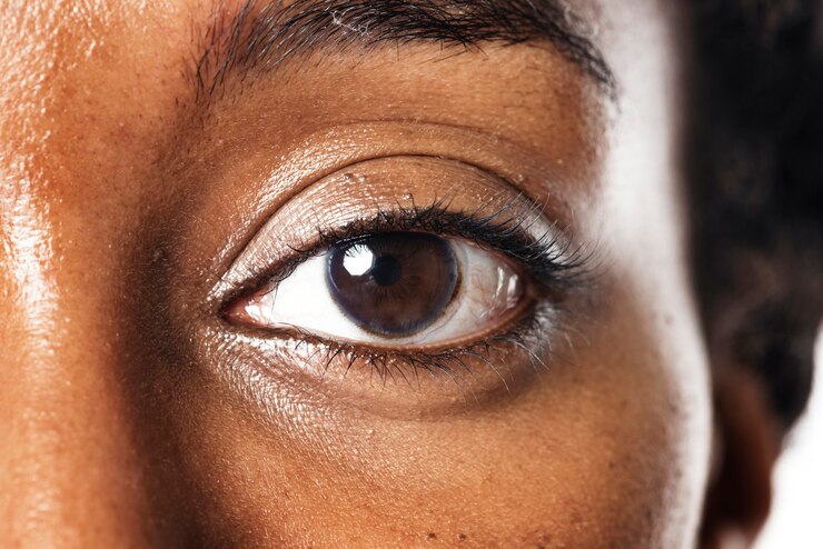
THYROID AND EYE HEALTH

The thyroid gland, a butterfly-shaped organ located in the neck, plays a crucial role in regulating metabolism and overall bodily functions. The thyroid gland produces hormones that regulate metabolism, energy levels, and various physiological processes. However, its influence extends beyond metabolic processes, significantly impacting eye health. When the thyroid gland malfunctions, it can lead to disorders such as hyperthyroidism (overactive thyroid) or hypothyroidism (underactive thyroid). One particular condition, Graves’ disease, an autoimmune disorder causing hyperthyroidism, is closely associated with eye problems, known as Thyroid Eye Disease (TED) or Graves’ orbitopathy.

THYROID EYE DISEASE (TED):

TED is an inflammatory condition affecting the muscles and tissues around the eyes. It is most commonly seen in individuals with Graves’ disease, although it can occasionally occur in those with hypothyroidism or even in people with normal thyroid function. TED can cause a range of symptoms from mild irritation to severe vision impairment, significantly impacting a person’s quality of life.

 HOW THYROID IMPACTS OUR VISION

Thyroid dysfunction can lead to several eye-related issues, primarily due to inflammation and abnormal immune responses. Here are the key ways thyroid problems can affect vision:

  1. Proptosis (Exophthalmos): One of the hallmark symptoms of TED is proptosis, where the eyes bulge forward. This occurs due to swelling and inflammation of the eye muscles and fatty tissues behind the eye. Proptosis can lead to a staring appearance and cause discomfort or pain.
  2. Dry Eyes: Thyroid dysfunction can reduce tear production, leading to dry eyes. Insufficient lubrication can cause irritation, redness, a gritty sensation, and even damage to the cornea over time.
  3. Double Vision (Diplopia): Swollen eye muscles can become stiff and misaligned, causing double vision. This can make daily activities like reading, driving, or even walking difficult and uncomfortable.
  4. Vision Loss: In severe cases, TED can compress the optic nerve, leading to optic neuropathy. This compression can result in vision loss, which may be irreversible if not treated promptly.
  5. Lid Retraction and Lag: The upper eyelid may retract (pull back) or lag behind when looking down, causing the eyes to appear more open than usual. This can contribute to dry eyes and increased sensitivity to light.

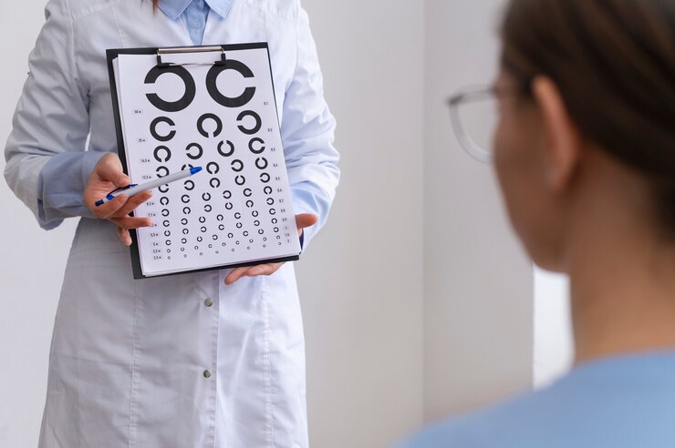
 SYMPTOMS TO IDENTIFY WHETHER THE THYROID IS DAMAGING YOUR EYES

Recognizing the symptoms of thyroid-related eye problems early is crucial for seeking appropriate treatment and preventing further damage. Here are common signs to watch for:

  1. Bulging Eyes: A noticeable bulging or protrusion of one or both eyes is a clear indicator of TED. This symptom can develop gradually and may be accompanied by discomfort or pain.
  2. Eye Redness and Irritation: Chronic redness, swelling, or a feeling of grittiness in the eyes can suggest thyroid-related inflammation. This is often due to dry eyes or irritation from swollen tissues.
  3. Excessive Tearing or Dryness: Paradoxically, thyroid eye disease can cause both excessive tearing and dry eyes. This imbalance occurs because of the disruption in normal tear production and drainage.
  4. Double Vision: Persistent double vision or difficulty focusing on objects can be a sign of misaligned eye muscles. This symptom can vary in severity and may worsen over time.
  5. Light Sensitivity: Increased sensitivity to light (photophobia) can occur due to eyelid retraction or corneal exposure from dry eyes. Bright lights may cause discomfort or pain.
  6. Difficulty Moving Eyes: Stiffness or pain when moving the eyes, particularly when looking up or sideways, can indicate inflamed or swollen eye muscles.
  7. Vision Changes: Blurred vision, reduced color perception, or sudden loss of vision are serious symptoms that require immediate medical attention. These changes could indicate optic nerve involvement.

 MANAGING THYROID-RELATED EYE PROBLEMS

Effective management of thyroid-related eye problems involves a combination of treating the underlying thyroid disorder and addressing the eye symptoms directly. Here are key strategies for managing these issues:

  1. Thyroid Treatment: Regulating thyroid hormone levels is the first step in managing TED. This may involve medications to control hyperthyroidism, radioactive iodine therapy, or thyroid surgery. Proper thyroid management can help reduce the severity of eye symptoms.
  2. Eye Lubrication: Using artificial tears or lubricating eye drops can help alleviate dryness and irritation. Gel or ointment formulations may be recommended for more severe cases, particularly at night.
  3. Steroid Therapy: Inflammatory symptoms may be treated with corticosteroids to reduce swelling and pain. These can be administered orally or intravenously, depending on the severity of the symptoms.
  4. Radiation Therapy: In some cases, low-dose radiation therapy may be used to reduce inflammation and swelling around the eyes. This treatment is typically considered when steroid therapy is insufficient.
  5. Surgical Intervention: Severe cases of TED may require surgical intervention to correct proptosis, relieve optic nerve compression, or improve eyelid function. Orbital decompression surgery, strabismus surgery (to correct double vision), and eyelid surgery are potential options.
  6. Lifestyle Modifications: Simple changes such as wearing sunglasses to reduce light sensitivity, using a humidifier to maintain moisture in the air, and applying cool compresses to reduce swelling can provide symptomatic relief.

Thyroid dysfunction can significantly impact eye health, particularly in conditions like Graves’ disease and thyroid eye disease. Understanding the connection between thyroid problems and vision is essential for early detection and effective management. Recognizing symptoms such as bulging eyes, double vision, and dry eyes can help identify thyroid-related eye issues. Comprehensive treatment, including thyroid regulation, eye lubrication, and potentially surgical interventions, is crucial for preserving vision and improving quality of life. If you experience any symptoms of thyroid-related eye problems, seek medical advice promptly to ensure timely and effective care.

For any eye related problems, schedule an appointment with Dr. Rani Menon Maxivision Eye Hospitals. We help you with latest technology and expert doctors.


Tags :
Blog
Share This :
Categories
Blog squint

Everything You Need to Know About Strabismus (Squint) Eye Surgery

Everything You Need to Know About Strabismus (Squint) Eye Surgery

squint-eye-surgery

Strabismus, commonly known as a squint, is a condition where the eyes do not align properly. One eye may turn in, out, up, or down while the other eye focuses on a single point. This misalignment can be constant or intermittent and can affect both children and adults. Strabismus surgery aims to correct this misalignment, improving both the appearance and functionality of the eyes. The surgery involves adjusting the muscles around the eyes to ensure they work together properly. This comprehensive guide provides detailed insights into strabismus, including its definition, affected demographics, symptoms, causes, and the surgical process. Understanding these aspects is crucial for those considering or preparing for strabismus surgery.

 WHAT IS STRABISMUS OR (SQUINT)?

Strabismus, or squint, is a visual disorder where the eyes do not look in the same direction at the same time. Normally, the brain coordinates the movement of the eyes so they point in the same direction and perceive depth accurately. However, in strabismus, there is a lack of coordination between the extraocular muscles, causing the eyes to deviate. This misalignment can be in any direction: inward (esotropia), outward (exotropia), upward (hypertropia), or downward (hypotropia). Strabismus can occur intermittently or constantly and may affect one or both eyes. It is essential to address strabismus promptly, as it can lead to amblyopia (lazy eye) where the brain starts ignoring the input from the misaligned eye, potentially resulting in permanent vision loss.

Strabismus can develop due to various reasons, including congenital factors, developmental issues, or acquired conditions. Congenital strabismus is often present at birth or develops in early infancy. Developmental strabismus can emerge as a child grows, often becoming noticeable between ages 2 and 4. Acquired strabismus can occur later in life due to trauma, medical conditions like diabetes or stroke, or other eye disorders. Diagnosing strabismus typically involves a comprehensive eye examination, including visual acuity tests, alignment assessments, and possibly imaging studies. Early detection and treatment are vital for preventing long-term vision problems and enhancing the quality of life for those affected.

WHO ALL ARE AFFECTED?

Strabismus can affect individuals of all ages, from infants to adults. However, it is most commonly diagnosed in children. Approximately 2-4% of the population is affected by strabismus, making it a relatively common eye disorder. In children, the condition often becomes noticeable during infancy or early childhood. Genetic factors can play a significant role, as a family history of strabismus increases the likelihood of developing the condition. Children with conditions such as Down syndrome or cerebral palsy are also at a higher risk.

IN ADULTS

Strabismus can develop due to various reasons, including untreated childhood strabismus that reoccurs or new onset due to medical conditions such as thyroid eye disease, diabetes, stroke, or neurological disorders. Trauma to the eye or head can also cause strabismus in adults. Additionally, certain surgeries or illnesses that affect the nervous system can lead to the development of strabismus.

IN CHILDREN

The impact of strabismus extends beyond physical misalignment of the eyes. In children, it can affect visual development, leading to amblyopia if not treated promptly. Social and psychological effects are also significant, as noticeable eye misalignment can lead to self-consciousness and social stigma. Adults with strabismus may face challenges in professional and social settings, affecting their quality of life. The comprehensive nature of strabismus underscores the importance of early detection, ongoing monitoring, and appropriate intervention to mitigate its impacts across all age groups.

 SYMPTOMS OF STRABISMUS

MISALIGNMENT OF THE EYES: The most noticeable symptom is the visible misalignment, where one eye may point in a different direction than the other.

DOUBLE VISION: Adults and older children with strabismus might experience double vision, as the brain receives two different images from each eye.

UNCOORDINATED EYE MOVEMENTS: The eyes may not move together smoothly. One eye might seem to lag behind or move independently.

HEAD TILTING OR TURNING: Individuals often tilt or turn their head to try to use both eyes together and reduce double vision or discomfort.

SQUINTING OR CLOSING ONE EYE: To improve vision or reduce double vision, a person might frequently squint or close one eye, especially in bright light or when focusing on distant objects.

DIFFICULTY WITH DEPTH PERCEPTION: Strabismus can impair depth perception, making it challenging to judge distances accurately.

EYE STRAIN AND FATIGUE: Strabismus can cause significant eye strain and fatigue, especially during tasks that require focused vision like reading or using a computer.

FREQUENT BLINKING OR EYE RUBBING: Children with strabismus may often blink excessively or rub their eyes, indicating discomfort or visual disturbance.

IN CHILDREN

The early symptoms can be subtle. Parents might notice their child’s eyes occasionally crossing or drifting. In some cases, the child may not show obvious symptoms but could have difficulties with reading or hand-eye coordination. Regular eye examinations are crucial, as early detection and treatment of strabismus can prevent complications such as amblyopia and enhance visual development. In adults, any sudden onset of strabismus requires immediate medical attention, as it could indicate an underlying health issue that needs prompt treatment.

CAUSES OF STRABISMUS

Genetic Factors: A family history of strabismus significantly increases the risk. Genetic predisposition can affect the development and function of the eye muscles and nerves.

Congenital Conditions: Strabismus present at birth or developing in infancy is often linked to congenital issues. Conditions like congenital esotropia or exotropia are relatively common in newborns.

Neurological Disorders: Problems with the brain or nervous system, such as cerebral palsy, Down syndrome, or hydrocephalus, can disrupt the coordination of eye movements, leading to strabismus.

Refractive Errors: Significant differences in vision between the two eyes (anisometropia) or severe nearsightedness, farsightedness, or astigmatism can cause the eyes to turn inwards or outwards.

Muscle Abnormalities: Dysfunction or malformation of the eye muscles can prevent proper alignment. This can be due to developmental issues or acquired conditions.

Medical Conditions: Diseases such as diabetes, thyroid eye disease, or stroke can affect the muscles or nerves controlling eye movement, leading to strabismus.

Injuries: Trauma to the eye or head can damage the muscles or nerves, resulting in misalignment.

Infections: Severe infections that affect the eye or brain, such as meningitis, can cause strabismus.

ABOUT STRABISMUS (SQUINT) EYE SURGERY

Strabismus eye surgery aims to correct the misalignment of the eyes by adjusting the muscles responsible for eye movement. This surgical intervention is typically considered when non-surgical treatments, such as glasses, prisms, or vision therapy, are insufficient in correcting the condition. The surgery can be performed on one or both eyes, depending on the specific needs of the patient.

Preoperative Assessment: A thorough preoperative evaluation is essential to determine the type and extent of muscle adjustment required. This assessment includes measuring the degree of misalignment, assessing the health and function of the eye muscles, and considering any underlying conditions contributing to the strabismus.

Surgical Procedure: During the surgery, which is usually performed under general anesthesia, the surgeon makes a small incision in the conjunctiva (the clear membrane covering the white part of the eye) to access the eye muscles. The muscles are then repositioned by either shortening (resecting) or lengthening (recessing) them, depending on whether the eye needs to be strengthened or weakened. The goal is to achieve proper alignment so that the eyes can work together more effectively.

Recovery: Postoperative care is crucial for a successful outcome. Patients may experience mild discomfort, redness, or swelling in the operated eye(s) for a few days. Eye drops or ointments are often prescribed to prevent infection and reduce inflammation. Follow-up visits are necessary to monitor the healing process and ensure that the eyes remain properly aligned.

Outcomes and Complications: The success rate of strabismus surgery is generally high, with many patients achieving significant improvement in eye alignment and binocular vision. However, some patients may require additional surgeries to fine-tune the alignment. Potential complications, though rare, include infection, bleeding, or adverse reactions to anesthesia. In some cases, overcorrection or under-correction of the alignment may occur, necessitating further treatment.

CONCLUSION

Strabismus, or squint, is a common eye disorder characterized by misaligned eyes, which can significantly impact visual development and quality of life. It affects individuals across all age groups, with varying symptoms such as double vision, eye strain, and impaired depth perception. The causes of strabismus are diverse, including genetic factors, congenital conditions, neurological disorders, refractive errors, and injuries. Strabismus eye surgery plays a crucial role in correcting eye alignment when other treatments are insufficient.

Schedule an appointment with Dr. Rani Menon Maxivision Eye Hospitals for the best eye care treatments in Kerala.

Tags :
Blog,squint
Share This :
Categories
Blog

Warning Signs of Retinal Detachment And What You Should Do

Warning Signs of Retinal Detachment And What You Should Do

retina-treatments-thrisur

UNDERSTANDING RETINAL DETACHMENT

Retinal detachment is a serious eye condition where the retina, a thin layer of tissue at the back of the eye, peels away from its underlying supportive tissue. This separation disrupts the retina’s normal functioning, leading to potential vision loss if not promptly treated. Understanding the causes, symptoms, diagnosis, and treatment options is crucial for effective management and prevention of this sight-threatening condition.


CAUSES AND RISK FACTORS

The primary cause of retinal detachment is the presence of a tear or hole in the retina, which allows fluid to seep underneath, separating it from the underlying tissues.

Rhegmatogenous Retinal Detachment: This is the most common type and is caused by a tear or break in the retina. Ageing is a significant risk factor because the vitreous, a gel-like substance inside the eye, shrinks and can pull on the retina, leading to tears. Other risk factors include severe myopia (near sightedness), previous eye surgery, or trauma.

Tractional Retinal Detachment: This occurs when scar tissue on the retina’s surface contracts, pulling the retina away from the back of the eye. It is often seen in people with diabetes mellitus, which can lead to diabetic retinopathy, a condition where abnormal blood vessels grow on the retina’s surface.

Exudative Retinal Detachment: This type is caused by inflammation, injury, or vascular abnormalities that result in fluid accumulation under the retina without any tears or breaks. Conditions such as inflammatory disorders, tumours, or age-related macular degeneration can lead to this type.

WARNING SIGNS OF RETINAL DETACHMENT

Retinal detachment is a serious condition that can lead to permanent vision loss if not promptly treated. Recognizing the warning signs early is crucial for seeking timely medical intervention. Here are the primary warning signs to watch out for:

Sudden Appearance of Floaters

Floaters are small specks or threads that drift through your field of vision. While floaters are common and usually benign, a sudden increase in their number can be a warning sign of retinal detachment. These floaters are caused by tiny clumps of gel or cells inside the vitreous, the clear gel-like substance that fills the inside of your eye.

Flashes of Light

Experiencing sudden flashes of light, particularly in your peripheral vision, is another common warning sign. These flashes can resemble lightning streaks and occur due to the vitreous pulling on the retina. The sensation can be more noticeable in dark environments.

Blurred Vision

A sudden decrease in vision clarity or blurring of vision can indicate retinal detachment. This blurriness often occurs in just one eye and can affect any part of your visual field.

Shadow or Curtain Over Vision

One of the most serious warning signs is the perception of a shadow or curtain descending over your field of vision. This effect can start in a small area and spread across the vision field as the detachment progresses. It typically starts from the peripheral (side) vision and moves towards the central vision.

Loss Of Peripheral Vision

Noticing a reduction or loss of peripheral (side) vision is a significant warning sign. This can be experienced as a darkening or shadow moving inward from the edges of your vision.

If you experience any of these warning signs, it is crucial to seek medical attention immediately. Retinal detachment is a medical emergency, and prompt treatment is essential to preserve vision. Early diagnosis and treatment can significantly improve the outcome and prevent permanent vision loss. Regular eye examinations, especially if you are at higher risk due to factors like severe nearsightedness, previous eye injuries, or family history, can help in early detection and management.

WHAT WE SHOULD DO?

Seek Immediate Medical Attention: If you experience any of these symptoms, contact an eye care professional immediately.

Avoid Strenuous Activity: Refrain from heavy lifting or vigorous exercise until you are evaluated by a doctor.

Keep Calm and Stay Still: Try to stay calm and avoid moving your eyes excessively to prevent further damage.

Prepare for an Eye Exam: Be ready for a comprehensive eye examination, which may include tests like ophthalmoscopy, ultrasound, or optical coherence tomography (OCT).

Follow Medical Advice: Adhere strictly to the treatment plan provided by your eye care specialist, which may include surgery or other interventions to repair the retina.

DIAGNOSIS

An eye examination is essential for identifying retinal detachment. An ophthalmologist will perform several tests, including:

Diagnostic Procedures for Retinal Detachment

Detecting retinal detachment early is critical for effective treatment and preventing permanent vision loss. A comprehensive eye examination can identify signs of retinal detachment. The following diagnostic procedures are commonly used by ophthalmologists:

 Visual Acuity Test

A Visual Acuity Test measures how well you can see at various distances. During this test, you will be asked to read letters on a chart (commonly known as a Snellen chart) from a specified distance. Each eye is tested separately, with and without corrective lenses if you wear them. The test determines the smallest letters you can read on the chart, indicating the clarity and sharpness of your vision. A decline in visual acuity may signal an underlying issue, such as retinal detachment, especially if it occurs suddenly.

Dilated Eye Exam

A dilated eye exam involves the use of special eyedrops to widen (dilate) the pupils, allowing the doctor to get a better view of the retina and other structures at the back of the eye. After administering the drops, which take about 15 to 30 minutes to fully dilate the pupils, the ophthalmologist uses a magnifying lens to inspect the retina for any tears, holes, or areas of detachment. The dilation provides a more comprehensive view, making it easier to detect abnormalities that might not be visible with non-dilated pupils. This examination can also reveal other eye conditions, such as macular degeneration or diabetic retinopathy.

Ophthalmoscopy

Ophthalmoscopy is a diagnostic procedure utilized to inspect the rear portion of the eye, encompassing the retina, optic disc, and blood vessels. The doctor uses an ophthalmoscope, which is a handheld instrument equipped with a light and several lenses. The ophthalmoscope allows for a detailed examination of the retina. The procedure can be performed directly, with the doctor looking through the pupil, or indirectly, using a special lens held close to the eye. Indirect ophthalmoscopy, often performed with the aid of scleral depression (pressing on the sclera or white of the eye), provides a wider view of the retina, which is especially useful for detecting peripheral retinal tears or detachments.

Ultrasound Imaging

Ultrasound imaging of the eye, also known as ocular ultrasonography, is used when retinal detachment is difficult to visualize due to opacities like vitreous Hemorrhage (bleeding into the vitreous). This non-invasive test involves placing a small probe on the closed eyelid after applying a gel to facilitate sound wave transmission. The probe emits high-frequency sound waves that bounce off the internal structures of the eye and create detailed images of the retina and surrounding tissues. These images can help the ophthalmologist identify the location and extent of a retinal detachment, as well as other possible abnormalities such as tumours or foreign bodies within the eye. Ultrasound is particularly useful in emergency settings where a clear view of the retina is obstructed.

TREATMENT

Laser Surgery (Photocoagulation): A laser is used to seal the retinal tear by creating small burns around it, preventing fluid from passing through.

Cryopexy: Freezing is used to reattach the retina by creating a scar that helps secure the retina to the eye wall.

Pneumatic Retinopexy: A gas bubble is injected into the vitreous cavity to push the retina back into place.

Scleral Buckling: A piece of silicone material is sewn onto the sclera (white of the eye) to push the wall of the eye against the detached retina.

Vitrectomy: Removal of the vitreous gel to relieve traction on the retina and replace it with a gas bubble or silicone oil.

A specialized hospital like Dr. Rani Menon Maxivision Eye Hospitals in Thrissur provides hope and enhance the quality of life for those affected by retinal detachment. Schedule a consultation with our experts. Our compassionate team at Dr. Rani Menon Maxivision Eye Hospitals is here to assist with all your diabetes-related health concerns, tailored to the type of diabetes you have.

Tags :
Blog
Share This :
Categories
Blog

The Importance of Regular Eye Exams

The Importance of Regular Eye Exams

eye-exam-clinic

The eyes are one of the most vital organs in the human body, playing a crucial role in our daily lives and overall well-being. Vision is often considered the most dominant of the senses, providing us with the ability to perceive and interpret the world around us. The importance of eyes can be understood through various facets of human experience, including health, safety, communication, and quality of life. The eyes are indispensable to human life, affecting nearly every aspect of our existence. Protecting our vision through regular eye check-ups, proper nutrition, and eye safety practices is essential for maintaining the richness and fullness of life that our eyes provide.

Health and Safety:

Our eyes serve as primary tools for navigating our environment safely. They help us detect hazards, recognize faces, and interpret signs, all of which are essential for avoiding accidents and staying safe. Good vision is critical for tasks that require precision and coordination, such as driving, reading, and operating machinery. Without a clear vision, these activities become challenging and potentially dangerous.

Communication:

Eyes are fundamental in non-verbal communication, allowing us to express and perceive emotions. Eye contact can convey confidence, interest, and understanding, playing a significant role in social interactions. Through our eyes, we can read the emotional states of others, fostering empathy and deeper connections.

Quality of Life:

The ability to see and appreciate the beauty of the world significantly enhances our quality of life. From enjoying a colorful sunset to appreciating art, vision enriches our experiences and adds depth to our perceptions. Visual stimuli are crucial for learning and cognitive development, especially in children, where visual aids are often used in education to facilitate better understanding.

Mental and Emotional Well-being:

Vision impairment can lead to social isolation and decreased independence, impacting mental health. People with good vision are more likely to engage in social activities and lead active lifestyles, contributing to their overall happiness and well-being.

Medical Insight:

Eyes can also be indicators of general health. Conditions like diabetes, hypertension, and even certain neurological disorders can manifest in the eyes, allowing for early diagnosis and treatment. Regular eye examinations can thus be pivotal in maintaining not just ocular health but overall health.


THE IMPORTANCE OF REGULAR EYE EXAMS

Regular eye exams are a critical component of maintaining overall health and well-being. Eyes are not only the windows to the world but also windows to our general health. Regular eye check-ups can detect eye problems at their earliest stages, when they are most treatable, and can also uncover other health issues.

Early Detection of Eye Diseases

One of the primary reasons for regular eye exams is the early detection of eye diseases. Many serious eye conditions, such as glaucoma, macular degeneration, and diabetic retinopathy, often develop without noticeable symptoms in the early stages. By the time symptoms become apparent, significant, irreversible damage may have already occurred. Regular eye exams can identify these diseases early, allowing for timely intervention that can prevent or minimize vision loss.

Monitoring Vision Changes

Vision changes can occur gradually and might go unnoticed until they significantly impact daily activities. Regular eye exams can detect subtle changes in vision that you might not be aware of. For example, changes in refractive errors such as myopia (near sightedness), hyperopia (far sightedness), and astigmatism can be easily corrected with updated prescriptions for glasses or contact lenses. Keeping your prescription current ensures that you can see as clearly and comfortably as possible.

Identifying General Health Issues

The eyes can reveal a lot about overall health. Conditions such as diabetes, hypertension, high cholesterol, and even certain cancers can manifest in the eyes. Through a comprehensive eye exam, an optometrist or ophthalmologist can spot signs of these systemic diseases and refer you to appropriate healthcare professionals for further evaluation and treatment. For instance, diabetic retinopathy is a common complication of diabetes that can be detected through an eye exam, often before the patient is aware of their diabetic condition.

Children’s Eye Health

Regular eye exams are particularly important for children. Vision is essential for a child’s development and learning.  Undetected vision problems can lead to difficulties in school and can impact a child’s ability to reach developmental milestones. Early eye exams can catch issues such as amblyopia (lazy eye), strabismus (crossed eyes), and other refractive errors, ensuring that children have the visual skills they need to succeed academically and socially.

Age-Related Eye Conditions

As we grow older, the likelihood of developing eye conditions such as cataracts, glaucoma, and macular degeneration rises. Regular eye exams become increasingly important for older adults to monitor for these conditions. Early detection and management can help preserve vision and maintain quality of life.

Eye Safety and Preventive Care

Regular eye exams also provide an opportunity to discuss eye safety and preventive care. Eye care professionals can offer advice on protecting your eyes from injury, reducing eyestrain, and maintaining eye health through proper nutrition and lifestyle choices. For instance, they can recommend wearing protective eyewear during certain activities or using appropriate lighting and ergonomic setups to minimize eyestrain.

Conclusion

Regular eye exams are essential for maintaining good vision and overall health. They enable the early detection and treatment of eye diseases, monitor vision changes, and can uncover systemic health issues. For children, they are vital in ensuring proper visual development, and for older adults, they help manage age-related eye conditions. Additionally, regular exams offer a chance to receive valuable advice on eye safety and preventive care. Prioritizing kids is a simple yet powerful way to protect one of your most important senses and maintain your overall well-being.


If you are considering an eye check-up for yourself or for your loved ones, consult a professional eye specialist only. Once you have decided to proceed, schedule an initial consultation

Tags :
Blog
Share This :